
EPAS-1 rabbit pAb from ELK Biotechnology Read more ›

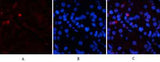
EPAS-1 rabbit pAb is a research-use antibody from ELK Biotechnology for studying EPAS-1 in WB, IHC, IF, ELISA workflows with listed reactivity for Human, Mouse, Rat. Specifications and supporting documents below should be reviewed when planning the experiment.
endothelial PAS domain protein 1(EPAS1) Homo sapiens This gene encodes a transcription factor involved in the induction of genes regulated by oxygen, which is induced as oxygen levels fall. The encoded protein contains a basic-helix-loop-helix domain protein dimerization domain as well as a domain found in proteins in signal transduction pathways which respond to oxygen levels. Mutations in this gene are associated with erythrocytosis familial type 4. [provided by RefSeq, Nov 2009],
| Specificity | EPAS-1 Polyclonal Antibody detects endogenous levels of EPAS-1 protein. |
|---|---|
| Validation evidence captions | Immunofluorescence analysis of human-stomach tissue. 1,EPAS-1 Polyclonal Antibody(red) was diluted at 1:200(4°C,overnight). 2, Cy3 labled Secondary antibody was diluted at 1:300(room temperature, 50min).3, Picture B: DAPI(blue) 10min. Picture A:Target. Picture B: DAPI. Picture C: merge of A+B | Immunofluorescence analysis of human-stomach tissue. 1,EPAS-1 Polyclonal Antibody(red) was diluted at 1:200(4°C,overnight). 2, Cy3 labled Secondary antibody was diluted at 1:300(room temperature, 50min).3, Picture B: DAPI(blue) 10min. Picture A:Target. Picture B: DAPI. Picture C: merge of A+B | Immunofluorescence analysis of mouse-spleen tissue. 1,EPAS-1 Polyclonal Antibody(red) was diluted at 1:200(4°C,overnight). 2, Cy3 labled Secondary antibody was diluted at 1:300(room temperature, 50min).3, Picture B: DAPI(blue) 10min. Picture A:Target. Picture B: DAPI. Picture C: merge of A+B | Immunofluorescence analysis of mouse-spleen tissue. 1,EPAS-1 Polyclonal Antibody(red) was diluted at 1:200(4°C,overnight). 2, Cy3 labled Secondary antibody was diluted at 1:300(room temperature, 50min).3, Picture B: DAPI(blue) 10min. Picture A:Target. Picture B: DAPI. Picture C: merge of A+B |
| Collaborator | ELK Biotechnology |
| Partner model | ES4055 |
| Target | EPAS-1 |
| Research areas | >>Pathways in cancer, >>Renal cell carcinoma |
| Reactivity | Human,Mouse,Rat |
| Applications | WB,IHC,IF,ELISA |
| Conjugation | Unconjugated |
| Dilution range | IF: 1:50-200 Western Blot: 1/500 - 1/2000. IHC-p: 1/100-1/300. ELISA: 1/20000. Not yet tested in other applications. |
| Host | Rabbit |
| Clonality | Polyclonal |
| Isotype | IgG |
| Immunogen | Synthesized peptide derived from human EPAS-1 around the non-acetylation site of K385. |
| Concentration | 1 mg/ml |
| Purification | The antibody was affinity-purified from rabbit antiserum by affinity-chromatography using epitope-specific immunogen. |
| Assay type | Antibody validation/application |
| Expression system | Expressed in most tissues, with highest levels in placenta, lung and heart. Selectively expressed in endothelial cells. |
| Purity | The antibody was affinity-purified from rabbit antiserum by affinity-chromatography using epitope-specific immunogen. |
| Species | WB | IHC | IF | ELISA | IP | FCM | ChIP |
|---|---|---|---|---|---|---|---|
| Human | |||||||
| Mouse | |||||||
| Rat |
| Species | Dilution | Notes |
|---|---|---|
| Human | IF: 1:50-200 Western Blot: 1/500 - 1/2000. IHC-p: 1/100-1/300. ELISA: 1/20000. Not yet tested in other applications. | — |
| Mouse | — | — |
| Rat | — | — |
| Species | Dilution | Notes |
|---|
| Species | Dilution | Notes |
|---|
| Species | Dilution | Notes |
|---|
Collaborator meets ABMIUM quality standards and overall product performance standards have been met.
Quality management certification.
For research applications. Not for diagnostic use.
No customer reviews yet.
