
NFATc4 rabbit pAb from ELK Biotechnology Read more ›

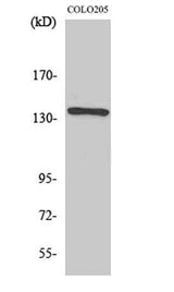
NFATc4 rabbit pAb is a research-use antibody from ELK Biotechnology for studying NFAT3 in WB, IHC, IF, ELISA workflows with listed reactivity for Human, Mouse. Specifications and supporting documents below should be reviewed when planning the experiment.
This gene encodes a member of the nuclear factor of activated T cells (NFAT) protein family. The encoded protein is part of a DNA-binding transcription complex. This complex consists of at least two components: a preexisting cytosolic component that translocates to the nucleus upon T cell receptor stimulation and an inducible nuclear component. NFAT proteins are activated by the calmodulin-dependent phosphatase, calcineurin. The encoded protein plays a role in the inducible expression of cytokine genes in T cells, especially in the induction of interleukin-2 and interleukin-4. Alternative splicing results in multiple transcript variants. [provided by RefSeq, Jan 2014],
| Specificity | NFATc4 Polyclonal Antibody detects endogenous levels of NFATc4 protein. |
|---|---|
| Validation evidence captions | Western Blot analysis of various cells using NFATc4 Polyclonal Antibody diluted at 1:500 | Immunofluorescence analysis of HeLa cells, using NFAT3 Antibody. The picture on the right is blocked with the synthesized peptide. | Immunohistochemistry analysis of paraffin-embedded human breast carcinoma tissue, using NFAT3 Antibody. The picture on the right is blocked with the synthesized peptide. | Western blot analysis of lysates from 293 cells, using NFAT3 Antibody. The lane on the right is blocked with the synthesized peptide. |
| Collaborator | ELK Biotechnology |
| Partner model | ES2938 |
| Target | NFAT3 |
| Research areas | >>cGMP-PKG signaling pathway, >>Cellular senescence, >>Wnt signaling pathway, >>Axon guidance, >>C-type lectin receptor signaling pathway, >>Oxytocin signaling pathway, >>Hepatitis B, >>Human cytomegalovirus infection, >>Human T-cell leukemia virus 1 infection, >>Kaposi sarcoma-associated herpesvirus infection, >>Human immunodeficiency virus 1 infection |
| Reactivity | Human,Mouse |
| Applications | WB,IHC,IF,ELISA |
| Conjugation | Unconjugated |
| Dilution range | Western Blot: 1/500 - 1/2000. Immunohistochemistry: 1/100 - 1/300. Immunofluorescence: 1/200 - 1/1000. ELISA: 1/10000. Not yet tested in other applications. |
| Host | Rabbit |
| Clonality | Polyclonal |
| Isotype | IgG |
| Immunogen | The antiserum was produced against synthesized peptide derived from human NFAT3. AA range:642-691 |
| Concentration | 1 mg/ml |
| Purification | The antibody was affinity-purified from rabbit antiserum by affinity-chromatography using epitope-specific immunogen. |
| Assay type | Antibody validation/application |
| Expression system | Widely expressed, with high levels in placenta, lung, kidney, testis and ovary (PubMed:18675896). Weakly expressed in spleen and thymus (PubMed:18675896). In the hippocampus, expressed in the granular layer of the dentate gyrus, in the pyramidal neurons of CA3 region, and in the hippocampal fissure (PubMed:18675896). Expressed in the heart (at protein level) (PubMed:12370307). |
| Purity | The antibody was affinity-purified from rabbit antiserum by affinity-chromatography using epitope-specific immunogen. |
| Species | WB | IHC | IF | ELISA | IP | FCM | ChIP |
|---|---|---|---|---|---|---|---|
| Human | |||||||
| Mouse |
| Species | Dilution | Notes |
|---|---|---|
| Human | Western Blot: 1/500 - 1/2000. Immunohistochemistry: 1/100 - 1/300. Immunofluorescence: 1/200 - 1/1000. ELISA: 1/10000. Not yet tested in other applications. | — |
| Mouse | — | — |
| Species | Dilution | Notes |
|---|
| Species | Dilution | Notes |
|---|
| Species | Dilution | Notes |
|---|
Collaborator meets ABMIUM quality standards and overall product performance standards have been met.
Quality management certification.
For research applications. Not for diagnostic use.
No customer reviews yet.
