OGT rabbit pAb
OGT rabbit pAb from ELK Biotechnology Read more ›

Product Description
OGT rabbit pAb is an ELK Biotechnology antibody for research workflows involving OGT.
Background
This gene encodes a glycosyltransferase that catalyzes the addition of a single N-acetylglucosamine in O-glycosidic linkage to serine or threonine residues. Since both phosphorylation and glycosylation compete for similar serine or threonine residues, the two processes may compete for sites, or they may alter the substrate specificity of nearby sites by steric or electrostatic effects. The protein contains multiple tetratricopeptide repeats that are required for optimal recognition of substrates. Alternatively spliced transcript variants encoding distinct isoforms have been found for this gene. [provided by RefSeq, Oct 2009],
Additional antibody information
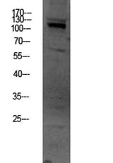
| Specificity | This antibody detects endogenous levels of OGT. |
|---|---|
| Validation evidence captions | Western blot analysis of HEPG2 lysate, antibody was diluted at 1000. Secondary antibody(catalog#:RS0002) was diluted at 1:20000 |
Key Facts
| Target | OGT |
| Also known as | UDP-N-acetylglucosamine--peptide N-acetylglucosaminyltransferase 110 kDa subunit (EC 2.4.1.255) (O-GlcNAc transferase subunit p110) (O-linked N-acetylglucosamine transferase 110 kDa subunit) (OGT) |
| Concentration | 1 mg/ml |
| Working concentration | WB 1:500-2000, ELISA 1:10000-20000 |
| Species reactivity | Human;Mouse;Rat |
| Observed band | 115kD |
| Cellular localization | Nucleus . Cytoplasm . Predominantly localizes to the nucleus. .; Isoform 2: Mitochondrion . Membrane . Associates with the mitochondrial inner membrane. .; Isoform 3: Cytoplasm . Nucleus . Cell membrane . Mitochondrion membrane . Cell projection . Mostly in the nucleus. Retained in the nucleus via interaction with HCFC1 (PubMed:21285374). After insulin induction, translocated from the nucleus to the cell membrane via phosphatidylinositide binding. Colocalizes with AKT1 at the plasma membrane. TRAK1 recruits this protein to mitochondria. In the absence of TRAK1, localizes in cytosol and nucleus (By similarity). .; Isoform 4: Cytoplasm. Nucleus. |
| Purity | The antibody was affinity-purified from rabbit antiserum by affinity-chromatography using epitope-specific immunogen. |
| Form / buffer | Liquid in PBS containing 50% glycerol, 0.5% BSA and 0.02% sodium azide. |
| Research area | >>Other types of O-glycan biosynthesis;>>Insulin resistance |
| Host species | Rabbit |
| Clonality | Polyclonal |
| Isotype | IgG |
| Conjugation | Unconjugated |
| Tested applications |
WBELISA |
| Dilution range | WB 1:500-2000, ELISA 1:10000-20000 |
| Immunogen | Synthesized peptide derived from human OGT Polyclonal AA range: 435-475 |
| Purification | The antibody was affinity-purified from rabbit antiserum by affinity-chromatography using epitope-specific immunogen. |
Reactivity & Application Validation
3 speciess| Species | WB | IHC | IF | ELISA | IP | FCM | CHIP |
|---|---|---|---|---|---|---|---|
| Human | |||||||
| Mouse | |||||||
| Rat |
| Species | Dilution | Notes |
|---|---|---|
| Human | WB 1:500-2000 | — |
| Mouse | — | — |
| Rat | — | — |
| Species | Dilution | Notes |
|---|
Specifications
Storage & Stability
Compliance & Certifications
Manufactured under ISO 9001:2015 quality management standards.
Not intended for diagnostic or therapeutic use.