Perforin 1 rabbit pAb
Perforin 1 rabbit pAb from ELK Biotechnology Read more ›

Product Description
Perforin 1 rabbit pAb is an ELK Biotechnology antibody for research workflows involving Perforin.
Background
The protein encoded by this gene has structural and functional similarities to complement component 9 (C9). Like C9, this protein creates transmembrane tubules and is capable of lysing non-specifically a variety of target cells. This protein is one of the main cytolytic proteins of cytolytic granules, and it is known to be a key effector molecule for T-cell- and natural killer-cell-mediated cytolysis. Defects in this gene cause familial hemophagocytic lymphohistiocytosis type 2 (HPLH2), a rare and lethal autosomal recessive disorder of early childhood. Alternative splicing results in multiple transcript variants encoding the same protein. [provided by RefSeq, Jul 2008],
Additional antibody information
| Specificity | Perforin 1 Polyclonal Antibody detects endogenous levels of Perforin 1 |
|---|---|
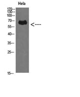
| Validation evidence captions | Western Blot analysis of Hela cells using Perforin 1 Polyclonal Antibody diluted at 1:500. Secondary antibody(catalog#:RS0002) was diluted at 1:20000 | Immunohistochemical analysis of paraffin-embedded human-kidney, antibody was diluted at 1:200 | Immunohistochemical analysis of paraffin-embedded human-kidney, antibody was diluted at 1:200 | Immunohistochemical analysis of paraffin-embedded human-spleen, antibody was diluted at 1:200 |
Key Facts
| Target | Perforin |
| Also known as | Perforin-1 (P1) (Cytolysin) (Lymphocyte pore-forming protein) (PFP) |
| Concentration | 1 mg/ml |
| Working concentration | WB 1:500-2000, IHC 1:50-200, ELISA 1:10000-20000 |
| Species reactivity | Human;Rat;Mouse; |
| Observed band | 61kD |
| Cellular localization | Cytolytic granule . Secreted. Cell membrane ; Multi-pass membrane protein . Endosome lumen . Stored in cytolytic granules of cytolytic T-lymphocytes and secreted into the cleft between T-lymphocyte and target cell (PubMed:20038786). Inserts into the cell membrane of target cells and forms pores (PubMed:20889983). Membrane insertion and pore formation requires a major conformation change (PubMed:20889983). May be taken up via endocytosis involving clathrin-coated vesicles and accumulate in a first time in large early endosomes (PubMed:20038786). . |
| Purity | The antibody was affinity-purified from rabbit antiserum by affinity-chromatography using epitope-specific immunogen. |
| Form / buffer | Liquid in PBS containing 50% glycerol, 0.5% BSA and 0.02% sodium azide. |
| Research area | >>Apoptosis;>>Natural killer cell mediated cytotoxicity;>>Type I diabetes mellitus;>>Autoimmune thyroid disease;>>Allograft rejection;>>Graft-versus-host disease;>>Viral myocarditis |
| Host species | Rabbit |
| Clonality | Polyclonal |
| Isotype | IgG |
| Conjugation | Unconjugated |
| Tested applications |
WBIHCELISA |
| Dilution range | WB 1:500-2000, IHC 1:50-200, ELISA 1:10000-20000 |
| Immunogen | The antiserum was produced against synthesized peptide derived from the C-terminal region of human PRF1. AA range:451-500 |
| Purification | The antibody was affinity-purified from rabbit antiserum by affinity-chromatography using epitope-specific immunogen. |
Reactivity & Application Validation
3 speciess| Species | WB | IHC | IF | ELISA | IP | FCM | CHIP |
|---|---|---|---|---|---|---|---|
| Human | |||||||
| Rat | |||||||
| Mouse |
| Species | Dilution | Notes |
|---|---|---|
| Human | WB 1:500-2000 | — |
| Rat | — | — |
| Mouse | — | — |
| Species | Dilution | Notes |
|---|
| Species | Dilution | Notes |
|---|
Specifications
Storage & Stability
Compliance & Certifications
Manufactured under ISO 9001:2015 quality management standards.
Not intended for diagnostic or therapeutic use.