ABM99806
Villin rabbit pAb
ISO 9001|ABMIUM Verified™|Antibody
Villin rabbit pAb from ELK Biotechnology Read more ›
Related formats

Zoom
Product Description
Villin rabbit pAb is an ELK Biotechnology antibody for research workflows involving Villin.
Background
This gene encodes a member of a family of calcium-regulated actin-binding proteins. This protein represents a dominant part of the brush border cytoskeleton which functions in the capping, severing, and bundling of actin filaments. Two mRNAs of 2.7 kb and 3.5 kb have been observed; they result from utilization of alternate poly-adenylation signals present in the terminal exon. [provided by RefSeq, Jul 2008],
Additional antibody information
| Specificity | Villin Polyclonal Antibody detects endogenous levels of Villin |
|---|---|
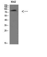
| Validation evidence captions | Western Blot analysis of K562 cells using Villin Polyclonal Antibody diluted at 1:500. Secondary antibody(catalog#:RS0002) was diluted at 1:20000 | Immunohistochemical analysis of paraffin-embedded human-colon, antibody was diluted at 1:200 | Immunohistochemical analysis of paraffin-embedded human-colon, antibody was diluted at 1:200 | Immunohistochemical analysis of paraffin-embedded human-kidney, antibody was diluted at 1:200 |
Key Facts
Product type
Primary Antibody›
Target: Villin
| Target | Villin |
| Also known as | villin 1 |
| Concentration | 1 mg/ml |
| Working concentration | WB 1:500-2000, ELISA 1:10000-20000 |
| Species reactivity | Human;Mouse;Rat |
| Observed band | 90kD |
| Cellular localization | Cytoplasm, cytoskeleton. Cell projection, lamellipodium. Cell projection, ruffle. Cell projection, microvillus. Cell projection, filopodium tip . Cell projection, filopodium . Relocalized in the tip of cellular protrusions and filipodial extensions upon infection with S.flexneri in primary intestinal epithelial cells (IEC) and in the tail-like structures forming the actin comets of S.flexneri. Redistributed to the leading edge of hepatocyte growth factor (HGF)-induced lamellipodia (By similarity). Rapidly redistributed to ruffles and lamellipodia structures in response to autotaxin, lysophosphatidic acid (LPA) and epidermal growth factor (EGF) treatment. . |
| Purity | The antibody was affinity-purified from rabbit antiserum by affinity-chromatography using epitope-specific immunogen. |
| Form / buffer | Liquid in PBS containing 50% glycerol, 0.5% BSA and 0.02% sodium azide. |
| Research area | Antibody Research |
| Host species | Rabbit |
| Clonality | Polyclonal |
| Isotype | IgG |
| Conjugation | Unconjugated |
| Tested applications |
WBELISA |
| Dilution range | WB 1:500-2000, ELISA 1:10000-20000 |
| Immunogen | Synthesized peptide derived from Villin at AA range: 601-650 |
| Purification | The antibody was affinity-purified from rabbit antiserum by affinity-chromatography using epitope-specific immunogen. |
Why researchers trust ABMIUM
2,000+
Researchers trust us
50,000+
Citations
ISO 9001
Certified Collaborators
Reactivity & Application Validation
3 speciess| Species | WB | IHC | IF | ELISA | IP | FCM | CHIP |
|---|---|---|---|---|---|---|---|
| Human | |||||||
| Mouse | |||||||
| Rat |
ABMIUM Validated™
WB
| Species | Dilution | Notes |
|---|---|---|
| Human | WB 1:500-2000 | — |
| Mouse | — | — |
| Rat | — | — |
ABMIUM Validated™
ELISA
| Species | Dilution | Notes |
|---|
Specifications
Target
Villin
Also known as
villin 1
Concentration
1 mg/ml
Working concentration
WB 1:500-2000, ELISA 1:10000-20000
Purity
The antibody was affinity-purified from rabbit antiserum by affinity-chromatography using epitope-specific immunogen.
Form / buffer
Liquid in PBS containing 50% glycerol, 0.5% BSA and 0.02% sodium azide.
Host species
Rabbit
Clonality
Polyclonal
Isotype
IgG
Conjugation
Unconjugated
Species reactivity
Human;Mouse;Rat
Tested applications
WBELISA
Immunogen
Synthesized peptide derived from Villin at AA range: 601-650
Observed band
90kD
Cellular localization
Cytoplasm, cytoskeleton. Cell projection, lamellipodium. Cell projection, ruffle. Cell projection, microvillus. Cell projection, filopodium tip . Cell projection, filopodium . Relocalized in the tip of cellular protrusions and filipodial extensions upon infection with S.flexneri in primary intestinal epithelial cells (IEC) and in the tail-like structures forming the actin comets of S.flexneri. Redistributed to the leading edge of hepatocyte growth factor (HGF)-induced lamellipodia (By similarity). Rapidly redistributed to ruffles and lamellipodia structures in response to autotaxin, lysophosphatidic acid (LPA) and epidermal growth factor (EGF) treatment. .
Dilution range
WB 1:500-2000, ELISA 1:10000-20000
Purification
The antibody was affinity-purified from rabbit antiserum by affinity-chromatography using epitope-specific immunogen.
Storage & Stability
Long-term storage
-20°C/1 year
Storage conditions
-20°C/1 year
Storage buffer
Liquid in PBS containing 50% glycerol, 0.5% BSA and 0.02% sodium azide.
Compliance & Certifications
ISO 9001
ISO 9001:2015 Certified
Manufactured under ISO 9001:2015 quality management standards.
RUO
For Research Use Only
Not intended for diagnostic or therapeutic use.
Customer Reviews
Recently Viewed