ABM99576
MAD1 rabbit pAb
ISO 9001|ABMIUM Verified™|Antibody
MAD1 rabbit pAb from ELK Biotechnology Read more ›
Related formats

Zoom
Product Description
MAD1 rabbit pAb is an ELK Biotechnology antibody for research workflows involving MAD1.
Background
MAD1L1 is a component of the mitotic spindle-assembly checkpoint that prevents the onset of anaphase until all chromosome are properly aligned at the metaphase plate. MAD1L1 functions as a homodimer and interacts with MAD2L1. MAD1L1 may play a role in cell cycle control and tumor suppression. Alternative splicing results in multiple transcript variants. [provided by RefSeq, Jan 2015],
Additional antibody information
| Specificity | MAD1 Polyclonal Antibody detects endogenous levels of MAD1 protein. |
|---|---|
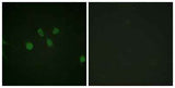
| Validation evidence captions | Immunofluorescence analysis of NIH/3T3 cells, using MAD1 Antibody. The picture on the right is blocked with the synthesized peptide. |
Key Facts
Product type
Primary Antibody›
Target: MAD1
| Target | MAD1 |
| Also known as | MAD1L1; MAD1; TXBP181; Mitotic spindle assembly checkpoint protein MAD1; Mitotic arrest deficient 1-like protein 1; MAD1-like protein 1; Mitotic checkpoint MAD1 protein homolog; HsMAD1; hMAD1; Tax-binding protein 181 |
| Concentration | 1 mg/ml |
| Working concentration | Immunofluorescence: 1/200 - 1/1000. ELISA: 1/10000. Not yet tested in other applications. |
| Species reactivity | Human;Rat;Mouse; |
| Cellular localization | Nucleus . Chromosome, centromere, kinetochore . Nucleus envelope . Cytoplasm, cytoskeleton, microtubule organizing center, centrosome . Cytoplasm, cytoskeleton, spindle . Cytoplasm, cytoskeleton, spindle pole . Co-localizes with TPR at the nucleus envelope during interphase and throughout the cell cycle (PubMed:22351768, PubMed:18981471). From the beginning to the end of mitosis, it is seen to move from a diffusely nuclear distribution to the centrosome, to the spindle midzone and finally to the midbody (PubMed:9546394). Localizes to kinetochores during prometaphase (PubMed:22351768, PubMed:29162720). Does not localize to kinetochores during metaphase (PubMed:29162720). Colocalizes with NEK2 at the kinetochore (PubMed:14978040). Colocalizes with IK at spindle poles during metaphase and ana |
| Purity | The antibody was affinity-purified from rabbit antiserum by affinity-chromatography using epitope-specific immunogen. |
| Form / buffer | Liquid in PBS containing 50% glycerol, 0.5% BSA and 0.02% sodium azide. |
| Research area | >>Cell cycle;>>Oocyte meiosis;>>Progesterone-mediated oocyte maturation;>>Human T-cell leukemia virus 1 infection;>>Viral carcinogenesis |
| Host species | Rabbit |
| Clonality | Polyclonal |
| Isotype | IgG |
| Conjugation | Unconjugated |
| Tested applications |
IFELISA |
| Dilution range | Immunofluorescence: 1/200 - 1/1000. ELISA: 1/10000. Not yet tested in other applications. |
| Immunogen | The antiserum was produced against synthesized peptide derived from human MAD1. AA range:394-443 |
| Purification | The antibody was affinity-purified from rabbit antiserum by affinity-chromatography using epitope-specific immunogen. |
Why researchers trust ABMIUM
2,000+
Researchers trust us
50,000+
Citations
ISO 9001
Certified Collaborators
Reactivity & Application Validation
3 speciess| Species | WB | IHC | IF | ELISA | IP | FCM | CHIP |
|---|---|---|---|---|---|---|---|
| Human | |||||||
| Rat | |||||||
| Mouse |
ABMIUM Validated™
IF
| Species | Dilution | Notes |
|---|---|---|
| Human | Immunofluorescence: 1/200 - 1/1000. ELISA: 1/10000. Not yet tested in other applications. | — |
| Rat | — | — |
| Mouse | — | — |
ABMIUM Validated™
ELISA
| Species | Dilution | Notes |
|---|
Specifications
Target
MAD1
Also known as
MAD1L1; MAD1; TXBP181; Mitotic spindle assembly checkpoint protein MAD1; Mitotic arrest deficient 1-like protein 1; MAD1-like protein 1; Mitotic checkpoint MAD1 protein homolog; HsMAD1; hMAD1; Tax-binding protein 181
Concentration
1 mg/ml
Working concentration
Immunofluorescence: 1/200 - 1/1000. ELISA: 1/10000. Not yet tested in other applications.
Purity
The antibody was affinity-purified from rabbit antiserum by affinity-chromatography using epitope-specific immunogen.
Form / buffer
Liquid in PBS containing 50% glycerol, 0.5% BSA and 0.02% sodium azide.
Host species
Rabbit
Clonality
Polyclonal
Isotype
IgG
Conjugation
Unconjugated
Species reactivity
Human;Rat;Mouse;
Tested applications
IFELISA
Immunogen
The antiserum was produced against synthesized peptide derived from human MAD1. AA range:394-443
Cellular localization
Nucleus . Chromosome, centromere, kinetochore . Nucleus envelope . Cytoplasm, cytoskeleton, microtubule organizing center, centrosome . Cytoplasm, cytoskeleton, spindle . Cytoplasm, cytoskeleton, spindle pole . Co-localizes with TPR at the nucleus envelope during interphase and throughout the cell cycle (PubMed:22351768, PubMed:18981471). From the beginning to the end of mitosis, it is seen to move from a diffusely nuclear distribution to the centrosome, to the spindle midzone and finally to the midbody (PubMed:9546394). Localizes to kinetochores during prometaphase (PubMed:22351768, PubMed:29162720). Does not localize to kinetochores during metaphase (PubMed:29162720). Colocalizes with NEK2 at the kinetochore (PubMed:14978040). Colocalizes with IK at spindle poles during metaphase and ana
Dilution range
Immunofluorescence: 1/200 - 1/1000. ELISA: 1/10000. Not yet tested in other applications.
Purification
The antibody was affinity-purified from rabbit antiserum by affinity-chromatography using epitope-specific immunogen.
Storage & Stability
Long-term storage
-20°C/1 year
Storage conditions
-20°C/1 year
Storage buffer
Liquid in PBS containing 50% glycerol, 0.5% BSA and 0.02% sodium azide.
Compliance & Certifications
ISO 9001
ISO 9001:2015 Certified
Manufactured under ISO 9001:2015 quality management standards.
RUO
For Research Use Only
Not intended for diagnostic or therapeutic use.
Customer Reviews
Recently Viewed