VCP (phospho Ser352) rabbit pAb
VCP (phospho Ser352) rabbit pAb from ELK Biotechnology Read more ›

Product Description
VCP (phospho Ser352) rabbit pAb is an ELK Biotechnology antibody for research workflows involving VCP.
Background
valosin containing protein(VCP) Homo sapiens The protein encoded by this gene is a member of a family that includes putative ATP-binding proteins involved in vesicle transport and fusion, 26S proteasome function, and assembly of peroxisomes. This protein, as a structural protein, is associated with clathrin, and heat-shock protein Hsc70, to form a complex. It has been implicated in a number of cellular events that are regulated during mitosis, including homotypic membrane fusion, spindle pole body function, and ubiquitin-dependent protein degradation. [provided by RefSeq, Jul 2008],
Additional antibody information
| Specificity | Phospho-VCP (S352) Polyclonal Antibody detects endogenous levels of VCP protein only when phosphorylated at S352. |
|---|---|
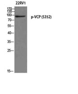
| Validation evidence captions | Western blot analysis of 22RV1 using p-VCP (S352) antibody. Antibody was diluted at 1:1000 | Western blot analysis of lysates from NIH/3T3 cells treated with starved 24h, using VCP (Phospho-Ser352) Antibody. The lane on the right is blocked with the phospho peptide. |
Key Facts
| Target | VCP |
| Also known as | VCP; Transitional endoplasmic reticulum ATPase; TER ATPase; 15S Mg(2+)-ATPase p97 subunit; Valosin-containing protein; VCP |
| Concentration | 1 mg/ml |
| Working concentration | Western Blot: 1/500 - 1/2000. ELISA: 1/5000. Not yet tested in other applications. |
| Species reactivity | Human;Mouse;Rat |
| Observed band | 85kD |
| Cellular localization | Cytoplasm, cytosol . Endoplasmic reticulum . Nucleus . Cytoplasm, Stress granule . Present in the neuronal hyaline inclusion bodies specifically found in motor neurons from amyotrophic lateral sclerosis patients (PubMed:15456787). Present in the Lewy bodies specifically found in neurons from Parkinson disease patients (PubMed:15456787). Recruited to the cytoplasmic surface of the endoplasmic reticulum via interaction with AMFR/gp78 (PubMed:16168377). Following DNA double-strand breaks, recruited to the sites of damage (PubMed:22120668). Recruited to stalled replication forks via interaction with SPRTN (PubMed:23042605). Recruited to damaged lysosomes decorated with K48-linked ubiquitin chains (PubMed:27753622). Colocalizes with TIA1, ZFAND1 and G3BP1 in cytoplasmic stress granules (SGs) in |
| Purity | The antibody was affinity-purified from rabbit antiserum by affinity-chromatography using epitope-specific immunogen. |
| Form / buffer | Liquid in PBS containing 50% glycerol, 0.5% BSA and 0.02% sodium azide. |
| Research area | >>Protein processing in endoplasmic reticulum;>>Amyotrophic lateral sclerosis;>>Pathways of neurodegeneration - multiple diseases;>>Legionellosis |
| Host species | Rabbit |
| Clonality | Polyclonal |
| Isotype | IgG |
| Conjugation | Unconjugated |
| Tested applications |
WBELISA |
| Dilution range | Western Blot: 1/500 - 1/2000. ELISA: 1/5000. Not yet tested in other applications. |
| Immunogen | The antiserum was produced against synthesized peptide derived from human VCP around the phosphorylation site of Ser352. AA range:318-367 |
| Purification | The antibody was affinity-purified from rabbit antiserum by affinity-chromatography using epitope-specific immunogen. |
Reactivity & Application Validation
3 speciess| Species | WB | IHC | IF | ELISA | IP | FCM | CHIP |
|---|---|---|---|---|---|---|---|
| Human | |||||||
| Mouse | |||||||
| Rat |
| Species | Dilution | Notes |
|---|---|---|
| Human | Western Blot: 1/500 - 1/2000. ELISA: 1/5000. Not yet tested in other applications. | — |
| Mouse | — | — |
| Rat | — | — |
| Species | Dilution | Notes |
|---|
Specifications
Storage & Stability
Compliance & Certifications
Manufactured under ISO 9001:2015 quality management standards.
Not intended for diagnostic or therapeutic use.