COP1 rabbit pAb
COP1 rabbit pAb from ELK Biotechnology Read more ›

Product Description
COP1 rabbit pAb is an ELK Biotechnology antibody for research workflows involving COP1.
Background
domain:The RING finger domain, in addition to its role in ubiquitination, functions as a structural scaffold to bring two clusters of positive-charged residues within spatial proximity to mimic a bipartite nuclear localization signal (NLS).,function:E3 ubiquitin-protein ligase that mediates ubiquitination and subsequent proteasomal degradation of target proteins. E3 ubiquitin ligases accept ubiquitin from an E2 ubiquitin-conjugating enzyme in the form of a thioester and then directly transfers the ubiquitin to targeted substrates. Involved in JUN ubiquitination and degradation. Directly involved in p53 (TP53) ubiquitination and degradation, thereby abolishing p53-dependent transcription and apoptosis. Ubiquitinates p53 independently of MDM2 or RCHY1. Probably mediates E3 ubiquitin ligase activity by functioning as the essential RING domain subunit of larger E3 complexes. In contrast, it does not constitute the catalytic RING subunit in the DCX DET1-COP1 complex that negatively regulates JUN, the ubiquitin ligase activity being mediated by RBX1.,induction:By p53/TP53.,pathway:Protein modification; protein ubiquitination.,similarity:Belongs to the COP1 family.,similarity:Contains 1 RING-type zinc finger.,similarity:Contains 7 WD repeats.,subcellular location:In the nucleus, it forms nuclear speckles.,subunit:Homodimer. Homodimerization is mediated by the coiled coil domain.
Additional antibody information
| Specificity | COP1 Polyclonal Antibody detects endogenous levels of COP1 protein. |
|---|---|
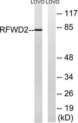
| Validation evidence captions | Western blot analysis of lysates from LOVO cells, using RFWD2 Antibody. The lane on the right is blocked with the synthesized peptide. | Western blot analysis of the lysates from HepG2 cells using RFWD2 antibody. |
Key Facts
| Target | COP1 |
| Also known as | RFWD2; COP1; RNF200; E3 ubiquitin-protein ligase RFWD2; Constitutive photomorphogenesis protein 1 homolog; hCOP1; RING finger and WD repeat domain protein 2; RING finger protein 200 |
| Concentration | 1 mg/ml |
| Working concentration | Western Blot: 1/500 - 1/2000. ELISA: 1/20000. Not yet tested in other applications. |
| Species reactivity | Human;Mouse |
| Observed band | 80kD |
| Cellular localization | Nucleus speckle. Cytoplasm. In the nucleus, it forms nuclear speckles. |
| Purity | The antibody was affinity-purified from rabbit antiserum by affinity-chromatography using epitope-specific immunogen. |
| Form / buffer | Liquid in PBS containing 50% glycerol, 0.5% BSA and 0.02% sodium azide. |
| Research area | >>p53 signaling pathway;>>Ubiquitin mediated proteolysis |
| Host species | Rabbit |
| Clonality | Polyclonal |
| Isotype | IgG |
| Conjugation | Unconjugated |
| Tested applications |
WBELISA |
| Dilution range | Western Blot: 1/500 - 1/2000. ELISA: 1/20000. Not yet tested in other applications. |
| Immunogen | The antiserum was produced against synthesized peptide derived from human RFWD2. AA range:661-710 |
| Purification | The antibody was affinity-purified from rabbit antiserum by affinity-chromatography using epitope-specific immunogen. |
Reactivity & Application Validation
2 speciess| Species | WB | IHC | IF | ELISA | IP | FCM | CHIP |
|---|---|---|---|---|---|---|---|
| Human | |||||||
| Mouse |
| Species | Dilution | Notes |
|---|---|---|
| Human | Western Blot: 1/500 - 1/2000. ELISA: 1/20000. Not yet tested in other applications. | — |
| Mouse | — | — |
| Species | Dilution | Notes |
|---|
Specifications
Storage & Stability
Compliance & Certifications
Manufactured under ISO 9001:2015 quality management standards.
Not intended for diagnostic or therapeutic use.