BAI-1 rabbit pAb
BAI-1 rabbit pAb from ELK Biotechnology Read more ›

Product Description
BAI-1 rabbit pAb is an ELK Biotechnology antibody for research workflows involving BAI-1.
Background
Angiogenesis is controlled by a local balance between stimulators and inhibitors of new vessel growth and is suppressed under normal physiologic conditions. Angiogenesis has been shown to be essential for growth and metastasis of solid tumors. In order to obtain blood supply for their growth, tumor cells are potently angiogenic and attract new vessels as results of increased secretion of inducers and decreased production of endogenous negative regulators. BAI1 contains at least one 'functional' p53-binding site within an intron, and its expression has been shown to be induced by wildtype p53. There are two other brain-specific angiogenesis inhibitor genes, designated BAI2 and BAI3 which along with BAI1 have similar tissue specificities and structures, however only BAI1 is transcriptionally regulated by p53. BAI1 is postulated to be a member of the secretin receptor family,
Additional antibody information
| Specificity | BAI-1 Polyclonal Antibody detects endogenous levels of BAI-1 protein. |
|---|---|
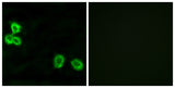
| Validation evidence captions | Immunofluorescence analysis of MCF7 cells, using BAI1 Antibody. The picture on the right is blocked with the synthesized peptide. | Immunohistochemistry analysis of paraffin-embedded human brain tissue, using BAI1 Antibody. The picture on the right is blocked with the synthesized peptide. | Western blot analysis of lysates from HepG2 cells, using BAI1 Antibody. The lane on the right is blocked with the synthesized peptide. | Western blot analysis of the lysates from HUVECcells using BAI1 antibody. |
Key Facts
| Target | BAI-1 |
| Also known as | BAI1; Brain-specific angiogenesis inhibitor 1 |
| Concentration | 1 mg/ml |
| Working concentration | Western Blot: 1/500 - 1/2000. Immunohistochemistry: 1/100 - 1/300. Immunofluorescence: 1/200 - 1/1000. ELISA: 1/10000. Not yet tested in other applications. |
| Species reactivity | Human;Mouse |
| Observed band | 174kD |
| Cellular localization | Cell membrane ; Multi-pass membrane protein . Cell projection, phagocytic cup . Cell junction, focal adhesion . Cell projection, dendritic spine . Cell junction, synapse, postsynaptic density .; Vasculostatin-120: Secreted .; Vasculostatin-40: Secreted . |
| Purity | The antibody was affinity-purified from rabbit antiserum by affinity-chromatography using epitope-specific immunogen. |
| Form / buffer | Liquid in PBS containing 50% glycerol, 0.5% BSA and 0.02% sodium azide. |
| Research area | >>p53 signaling pathway |
| Host species | Rabbit |
| Clonality | Polyclonal |
| Isotype | IgG |
| Conjugation | Unconjugated |
| Tested applications |
WBIHCIFELISA |
| Dilution range | Western Blot: 1/500 - 1/2000. Immunohistochemistry: 1/100 - 1/300. Immunofluorescence: 1/200 - 1/1000. ELISA: 1/10000. Not yet tested in other applications. |
| Immunogen | The antiserum was produced against synthesized peptide derived from human BAI1. AA range:691-740 |
| Purification | The antibody was affinity-purified from rabbit antiserum by affinity-chromatography using epitope-specific immunogen. |
Reactivity & Application Validation
2 speciess| Species | WB | IHC | IF | ELISA | IP | FCM | CHIP |
|---|---|---|---|---|---|---|---|
| Human | |||||||
| Mouse |
| Species | Dilution | Notes |
|---|---|---|
| Human | Western Blot: 1/500 - 1/2000. Immunohistochemistry: 1/100 - 1/300. Immunofluorescence: 1/200 - 1/1000. ELISA: 1/10000. Not yet tested in other applications. | — |
| Mouse | — | — |
| Species | Dilution | Notes |
|---|
| Species | Dilution | Notes |
|---|
| Species | Dilution | Notes |
|---|
Specifications
Storage & Stability
Compliance & Certifications
Manufactured under ISO 9001:2015 quality management standards.
Not intended for diagnostic or therapeutic use.