Septin 2 rabbit pAb
Septin 2 rabbit pAb from ELK Biotechnology Read more ›

Product Description
Septin 2 rabbit pAb is an ELK Biotechnology antibody for research workflows involving Septin 2.
Background
function:Required for normal progress through mitosis. Involved in cytokinesis.,similarity:Belongs to the septin family.,subcellular location:Accumulates near the contractile ring from anaphase through telophase, and finally condenses into the midbody. In interphase and postmitotic cells, localised to fibrous or granular structures, depending on the growth state of the cell.,subunit:Monomer, and homodimer. Nucleotide binding promotes dimerization. Heterohexamer composed of two heterotrimers containing one copy each of SEPT2, SEPT6 and SEPT7. The asymmetrical heterotrimers associate head-to-head to form a hexameric unit that assembles into filaments.,
Additional antibody information
| Specificity | Septin 2 Polyclonal Antibody detects endogenous levels of Septin 2 protein. |
|---|---|
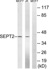
| Validation evidence captions | Western blot analysis of lysates from Jurkat and MCF-7 cells, using SEPT2 Antibody. The lane on the right is blocked with the synthesized peptide. | Western blot analysis of the lysates from HepG2 cells using SEPT2 antibody. | Immunohistochemical analysis of paraffin-embedded human Colon cancer. 1, Antibody was diluted at 1:200(4° overnight). 2, Tris-EDTA,pH9.0 was used for antigen retrieval. 3,Secondary antibody was diluted at 1:200(room temperature, 45min). |
Key Facts
| Target | Septin 2 |
| Also known as | SEPT2; DIFF6; KIAA0158; NEDD5; Septin-2; Neural precursor cell expressed developmentally down-regulated protein 5; NEDD-5 |
| Concentration | 1 mg/ml |
| Working concentration | WB 1:500-2000;IHC-p 1:50-300 |
| Species reactivity | Human;Mouse;Rat |
| Observed band | 41kD |
| Cellular localization | Cytoplasm . Cytoplasm, cytoskeleton . Cytoplasm, cytoskeleton, spindle . Chromosome, centromere, kinetochore . Cleavage furrow . Midbody . Cytoplasm, cell cortex . Cell projection, cilium membrane . Cell projection, cilium, flagellum . In metaphase cells, localized within the microtubule spindle. At the metaphase plate, in close apposition to the kinetochores of the congressed chromosomes. In cells undergoing cytokinesis, localized to the midbody, the ingressing cleavage furrow, and the central spindle. During bacterial infection, displays a collar shape structure next to actin at the pole of invading bacteria. In epithelial cells, colocalizes with polyglutamylated tubulin around the trans-Golgi network, as well as juxatnuclear and proximal Golgi apparatus. Localizes at the base of the cil |
| Purity | The antibody was affinity-purified from rabbit antiserum by affinity-chromatography using epitope-specific immunogen. |
| Form / buffer | Liquid in PBS containing 50% glycerol, 0.5% BSA and 0.02% sodium azide. |
| Research area | >>Bacterial invasion of epithelial cells;>>Shigellosis |
| Host species | Rabbit |
| Clonality | Polyclonal |
| Isotype | IgG |
| Conjugation | Unconjugated |
| Tested applications |
WBIHC |
| Dilution range | WB 1:500-2000;IHC-p 1:50-300 |
| Immunogen | The antiserum was produced against synthesized peptide derived from human SEPT2. AA range:103-152 |
| Purification | The antibody was affinity-purified from rabbit antiserum by affinity-chromatography using epitope-specific immunogen. |
Reactivity & Application Validation
3 speciess| Species | WB | IHC | IF | ELISA | IP | FCM | CHIP |
|---|---|---|---|---|---|---|---|
| Human | |||||||
| Mouse | |||||||
| Rat |
| Species | Dilution | Notes |
|---|---|---|
| Human | WB 1:500-2000;IHC-p 1:50-300 | — |
| Mouse | — | — |
| Rat | — | — |
| Species | Dilution | Notes |
|---|
Specifications
Storage & Stability
Compliance & Certifications
Manufactured under ISO 9001:2015 quality management standards.
Not intended for diagnostic or therapeutic use.