Lck rabbit pAb
Lck rabbit pAb from ELK Biotechnology Read more ›

Product Description
Lck rabbit pAb is an ELK Biotechnology antibody for research workflows involving Lck.
Background
This gene is a member of the Src family of protein tyrosine kinases (PTKs). The encoded protein is a key signaling molecule in the selection and maturation of developing T-cells. It contains N-terminal sites for myristylation and palmitylation, a PTK domain, and SH2 and SH3 domains which are involved in mediating protein-protein interactions with phosphotyrosine-containing and proline-rich motifs, respectively. The protein localizes to the plasma membrane and pericentrosomal vesicles, and binds to cell surface receptors, including CD4 and CD8, and other signaling molecules. Multiple alternatively spliced variants encoding different isoforms have been described. [provided by RefSeq, Aug 2016],
Additional antibody information
| Specificity | Lck Polyclonal Antibody detects endogenous levels of Lck protein. |
|---|---|
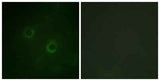
| Validation evidence captions | Immunofluorescence analysis of COS7 cells, using Lck Antibody. The picture on the right is blocked with the synthesized peptide. | Western blot analysis of lysates from Jurkat cells, using Lck Antibody. The lane on the right is blocked with the synthesized peptide. | Immunohistochemical analysis of paraffin-embedded human tonsil. 1, Antibody was diluted at 1:200(4° overnight). 2, Tris-EDTA,pH9.0 was used for antigen retrieval. 3,Secondary antibody was diluted at 1:200(room temperature, 45min). |
Key Facts
| Target | Lck |
| Also known as | LCK; Tyrosine-protein kinase Lck; Leukocyte C-terminal Src kinase; LSK; Lymphocyte cell-specific protein-tyrosine kinase; Protein YT16; Proto-oncogene Lck; T cell-specific protein-tyrosine kinase; p56-LCK |
| Concentration | 1 mg/ml |
| Working concentration | Western Blot: 1/500 - 1/2000. Immunohistochemistry: 1/100 - 1/300. Immunofluorescence: 1/200 - 1/1000. ELISA: 1/5000. Not yet tested in other applications. |
| Species reactivity | Human;Mouse;Rat |
| Observed band | 56kD |
| Cellular localization | Cell membrane ; Lipid-anchor ; Cytoplasmic side . Cytoplasm, cytosol . Present in lipid rafts in an inactive form. . |
| Purity | The antibody was affinity-purified from rabbit antiserum by affinity-chromatography using epitope-specific immunogen. |
| Form / buffer | Liquid in PBS containing 50% glycerol, 0.5% BSA and 0.02% sodium azide. |
| Research area | >>NF-kappa B signaling pathway;>>Osteoclast differentiation;>>Natural killer cell mediated cytotoxicity;>>Th1 and Th2 cell differentiation;>>Th17 cell differentiation;>>T cell receptor signaling pathway;>>Yersinia infection;>>Human T-cell leukemia virus 1 infection;>>PD-L1 expression and PD-1 checkpoint pathway in cancer;>>Primary immunodeficiency |
| Host species | Rabbit |
| Clonality | Polyclonal |
| Isotype | IgG |
| Conjugation | Unconjugated |
| Tested applications |
WBIHCIFELISA |
| Dilution range | Western Blot: 1/500 - 1/2000. Immunohistochemistry: 1/100 - 1/300. Immunofluorescence: 1/200 - 1/1000. ELISA: 1/5000. Not yet tested in other applications. |
| Immunogen | The antiserum was produced against synthesized peptide derived from human Lck. AA range:161-210 |
| Purification | The antibody was affinity-purified from rabbit antiserum by affinity-chromatography using epitope-specific immunogen. |
Reactivity & Application Validation
3 speciess| Species | WB | IHC | IF | ELISA | IP | FCM | CHIP |
|---|---|---|---|---|---|---|---|
| Human | |||||||
| Mouse | |||||||
| Rat |
| Species | Dilution | Notes |
|---|---|---|
| Human | Western Blot: 1/500 - 1/2000. Immunohistochemistry: 1/100 - 1/300. Immunofluorescence: 1/200 - 1/1000. ELISA: 1/5000. Not yet tested in other applications. | — |
| Mouse | — | — |
| Rat | — | — |
| Species | Dilution | Notes |
|---|
| Species | Dilution | Notes |
|---|
| Species | Dilution | Notes |
|---|
Specifications
Storage & Stability
Compliance & Certifications
Manufactured under ISO 9001:2015 quality management standards.
Not intended for diagnostic or therapeutic use.