ABM98658
TPX2 rabbit pAb
ISO 9001|ABMIUM Verified™|Antibody
TPX2 rabbit pAb from ELK Biotechnology Read more ›
Related formats

Zoom
Product Description
TPX2 rabbit pAb is an ELK Biotechnology antibody for research workflows involving TPX2.
Background
developmental stage:Exclusively expressed in proliferating cells from the transition G1/S until the end of cytokinesis.,PTM:Phosphorylated upon DNA damage, probably by ATM or ATR.,subcellular location:During mitosis it is strictly associated with the spindle pole and with the mitotic spindle, whereas during S and G2, it is diffusely distributed throughout the nucleus.,tissue specificity:Expressed in lung carcinoma cell lines but not in normal lung tissues.,
Additional antibody information
| Specificity | TPX2 Polyclonal Antibody detects endogenous levels of TPX2 protein. |
|---|---|
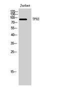
| Validation evidence captions | Western Blot analysis of Jurkat cells using TPX2 Polyclonal Antibody. Secondary antibody(catalog#:RS0002) was diluted at 1:20000 cells nucleus extracted by Minute TM Cytoplasmic and Nuclear Fractionation kit (SC-003,Inventbiotech,MN,USA). | Immunohistochemistry analysis of paraffin-embedded human brain tissue, using DIL-2 Antibody. The picture on the right is blocked with the synthesized peptide. | Western blot analysis of lysates from Jurkat cells, using DIL-2 Antibody. The lane on the right is blocked with the synthesized peptide. | Western blot analysis of the lysates from HUVECcells using DIL-2 antibody. |
Key Facts
Product type
Primary Antibody›
Target: TPX2
| Target | TPX2 |
| Also known as | TPX2; C20orf1; C20orf2; DIL2; HCA519; Targeting protein for Xklp2; Differentially expressed in cancerous and non-cancerous lung cells 2; DIL-2; Hepatocellular carcinoma-associated antigen 519; Protein fls353; Restricted expression prolifera |
| Concentration | 1 mg/ml |
| Working concentration | Western Blot: 1/500 - 1/2000. Immunohistochemistry: 1/100 - 1/300. ELISA: 1/10000. Not yet tested in other applications. |
| Species reactivity | Human;Mouse |
| Observed band | 86kD |
| Cellular localization | Nucleus . Cytoplasm, cytoskeleton, spindle . Cytoplasm, cytoskeleton, spindle pole . During mitosis it is strictly associated with the spindle pole and with the mitotic spindle, whereas during S and G2, it is diffusely distributed throughout the nucleus. Is released from the nucleus in apoptotic cells and is detected on apoptotic microtubules. . |
| Purity | The antibody was affinity-purified from rabbit antiserum by affinity-chromatography using epitope-specific immunogen. |
| Form / buffer | Liquid in PBS containing 50% glycerol, 0.5% BSA and 0.02% sodium azide. |
| Research area | Antibody Research |
| Host species | Rabbit |
| Clonality | Polyclonal |
| Isotype | IgG |
| Conjugation | Unconjugated |
| Tested applications |
WBIHCIFELISA |
| Dilution range | Western Blot: 1/500 - 1/2000. Immunohistochemistry: 1/100 - 1/300. ELISA: 1/10000. Not yet tested in other applications. |
| Immunogen | The antiserum was produced against synthesized peptide derived from human DIL-2. AA range:301-350 |
| Purification | The antibody was affinity-purified from rabbit antiserum by affinity-chromatography using epitope-specific immunogen. |
Why researchers trust ABMIUM
2,000+
Researchers trust us
50,000+
Citations
ISO 9001
Certified Collaborators
Reactivity & Application Validation
2 speciess| Species | WB | IHC | IF | ELISA | IP | FCM | CHIP |
|---|---|---|---|---|---|---|---|
| Human | |||||||
| Mouse |
ABMIUM Validated™
WB
| Species | Dilution | Notes |
|---|---|---|
| Human | Western Blot: 1/500 - 1/2000. Immunohistochemistry: 1/100 - 1/300. ELISA: 1/10000. Not yet tested in other applications. | — |
| Mouse | — | — |
ABMIUM Validated™
IHC
| Species | Dilution | Notes |
|---|
ABMIUM Validated™
IF
| Species | Dilution | Notes |
|---|
ABMIUM Validated™
ELISA
| Species | Dilution | Notes |
|---|
Specifications
Target
TPX2
Also known as
TPX2; C20orf1; C20orf2; DIL2; HCA519; Targeting protein for Xklp2; Differentially expressed in cancerous and non-cancerous lung cells 2; DIL-2; Hepatocellular carcinoma-associated antigen 519; Protein fls353; Restricted expression prolifera
Concentration
1 mg/ml
Working concentration
Western Blot: 1/500 - 1/2000. Immunohistochemistry: 1/100 - 1/300. ELISA: 1/10000. Not yet tested in other applications.
Purity
The antibody was affinity-purified from rabbit antiserum by affinity-chromatography using epitope-specific immunogen.
Form / buffer
Liquid in PBS containing 50% glycerol, 0.5% BSA and 0.02% sodium azide.
Host species
Rabbit
Clonality
Polyclonal
Isotype
IgG
Conjugation
Unconjugated
Species reactivity
Human;Mouse
Tested applications
WBIHCIFELISA
Immunogen
The antiserum was produced against synthesized peptide derived from human DIL-2. AA range:301-350
Observed band
86kD
Cellular localization
Nucleus . Cytoplasm, cytoskeleton, spindle . Cytoplasm, cytoskeleton, spindle pole . During mitosis it is strictly associated with the spindle pole and with the mitotic spindle, whereas during S and G2, it is diffusely distributed throughout the nucleus. Is released from the nucleus in apoptotic cells and is detected on apoptotic microtubules. .
Dilution range
Western Blot: 1/500 - 1/2000. Immunohistochemistry: 1/100 - 1/300. ELISA: 1/10000. Not yet tested in other applications.
Purification
The antibody was affinity-purified from rabbit antiserum by affinity-chromatography using epitope-specific immunogen.
Storage & Stability
Long-term storage
-20°C/1 year
Storage conditions
-20°C/1 year
Storage buffer
Liquid in PBS containing 50% glycerol, 0.5% BSA and 0.02% sodium azide.
Compliance & Certifications
ISO 9001
ISO 9001:2015 Certified
Manufactured under ISO 9001:2015 quality management standards.
RUO
For Research Use Only
Not intended for diagnostic or therapeutic use.
Customer Reviews
Recently Viewed