ACK (phospho Tyr284) rabbit pAb
ACK (phospho Tyr284) rabbit pAb from ELK Biotechnology Read more ›

Product Description
ACK (phospho Tyr284) rabbit pAb is an ELK Biotechnology antibody for research workflows involving ACK.
Background
This gene encodes a tyrosine kinase that binds Cdc42Hs in its GTP-bound form and inhibits both the intrinsic and GTPase-activating protein (GAP)-stimulated GTPase activity of Cdc42Hs. This binding is mediated by a unique sequence of 47 amino acids C-terminal to an SH3 domain. The protein may be involved in a regulatory mechanism that sustains the GTP-bound active form of Cdc42Hs and which is directly linked to a tyrosine phosphorylation signal transduction pathway. Several alternatively spliced transcript variants have been identified from this gene, but the full-length nature of only two transcript variants has been determined. [provided by RefSeq, Jul 2008],
Additional antibody information
| Specificity | Phospho-ACK (Y284) Polyclonal Antibody detects endogenous levels of ACK protein only when phosphorylated at Y284. |
|---|---|
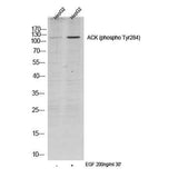
| Validation evidence captions | Western blot analysis of lysates from HepG2 cells, treated with EGF 200ng/ml 30', (Green) primary antibody was diluted at 1:1000, 4°over night, secondary antibody(cat:RS23920)was diluted at 1:10000, 37° 1hour. | Enzyme-Linked Immunosorbent Assay (Phospho-ELISA) for Immunogen Phosphopeptide (Phospho-left) and Non-Phosphopeptide (Phospho-right), using ACK1 (Phospho-Tyr284) Antibody | Immunofluorescence analysis of A549 cells, using ACK1 (Phospho-Tyr284) Antibody. The picture on the right is blocked with the phospho peptide. | Immunohistochemistry analysis of paraffin-embedded human breast carcinoma, using ACK1 (Phospho-Tyr284) Antibody. The picture on the right is blocked with the phospho peptide. |
Key Facts
| Target | ACK |
| Also known as | TNK2; ACK1; Activated CDC42 kinase 1; ACK-1; Tyrosine kinase non-receptor protein 2 |
| Concentration | 1 mg/ml |
| Working concentration | Western Blot: 1/500 - 1/2000. Immunohistochemistry: 1/100 - 1/300. Immunofluorescence: 1/200 - 1/1000. ELISA: 1/10000. Not yet tested in other applications. |
| Species reactivity | Human;Mouse |
| Observed band | 120kD |
| Cellular localization | Cell membrane . Nucleus . Endosome . Cell junction, adherens junction . Cytoplasmic vesicle membrane; Peripheral membrane protein; Cytoplasmic side . Cytoplasmic vesicle, clathrin-coated vesicle . Membrane, clathrin-coated pit . Cytoplasm, perinuclear region . Cytoplasm, cytosol . The Tyr-284 phosphorylated form is found both in the membrane and nucleus (By similarity). Co-localizes with EGFR on endosomes (PubMed:20333297). Nuclear translocation is CDC42-dependent (By similarity). Detected in long filamentous cytosolic structures where it co-localizes with CTPS1 (By similarity). . |
| Purity | The antibody was affinity-purified from rabbit antiserum by affinity-chromatography using epitope-specific immunogen. |
| Form / buffer | Liquid in PBS containing 50% glycerol, 0.5% BSA and 0.02% sodium azide. |
| Research area | Antibody Research |
| Host species | Rabbit |
| Clonality | Polyclonal |
| Isotype | IgG |
| Conjugation | Unconjugated |
| Tested applications |
WBIHCIFELISA |
| Dilution range | Western Blot: 1/500 - 1/2000. Immunohistochemistry: 1/100 - 1/300. Immunofluorescence: 1/200 - 1/1000. ELISA: 1/10000. Not yet tested in other applications. |
| Immunogen | The antiserum was produced against synthesized peptide derived from human ACK1 around the phosphorylation site of Tyr284. AA range:250-299 |
| Purification | The antibody was affinity-purified from rabbit antiserum by affinity-chromatography using epitope-specific immunogen. |
Reactivity & Application Validation
2 speciess| Species | WB | IHC | IF | ELISA | IP | FCM | CHIP |
|---|---|---|---|---|---|---|---|
| Human | |||||||
| Mouse |
| Species | Dilution | Notes |
|---|---|---|
| Human | Western Blot: 1/500 - 1/2000. Immunohistochemistry: 1/100 - 1/300. Immunofluorescence: 1/200 - 1/1000. ELISA: 1/10000. Not yet tested in other applications. | — |
| Mouse | — | — |
| Species | Dilution | Notes |
|---|
| Species | Dilution | Notes |
|---|
| Species | Dilution | Notes |
|---|
Specifications
Storage & Stability
Compliance & Certifications
Manufactured under ISO 9001:2015 quality management standards.
Not intended for diagnostic or therapeutic use.