CD98 rabbit pAb
CD98 rabbit pAb from ELK Biotechnology Read more ›

Product Description
CD98 rabbit pAb is an ELK Biotechnology antibody for research workflows involving CD98.
Background
This gene is a member of the solute carrier family and encodes a cell surface, transmembrane protein. The protein exists as the heavy chain of a heterodimer, covalently bound through di-sulfide bonds to one of several possible light chains. The encoded transporter plays a role in regulation of intracellular calcium levels and transports L-type amino acids. Alternatively spliced transcript variants, encoding different isoforms, have been characterized. [provided by RefSeq, Nov 2010],
Additional antibody information
| Specificity | CD98 Polyclonal Antibody detects endogenous levels of CD98 protein. |
|---|---|
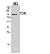
| Validation evidence captions | Western Blot analysis of HEB cells using CD98 Polyclonal Antibody. Secondary antibody(catalog#:RS0002) was diluted at 1:20000 |
Key Facts
| Target | CD98 |
| Also known as | SLC3A2; MDU1; 4F2 cell-surface antigen heavy chain; 4F2hc; 4F2 heavy chain antigen; Lymphocyte activation antigen 4F2 large subunit; CD98 |
| Concentration | 1 mg/ml |
| Working concentration | Western Blot: 1/500 - 1/2000. ELISA: 1/10000. Not yet tested in other applications. |
| Species reactivity | Human;Mouse;Rat |
| Observed band | 69kD |
| Cellular localization | Apical cell membrane . Cell membrane ; Single-pass type II membrane protein . Cell junction . Lysosome membrane . Melanosome . Localized at the plasma membrane when associated with SLC7A5/LAT1 or SLC7A8/LAT2 (PubMed:9751058, PubMed:11311135). Localized to the apical membrane of placental syncytiotrophoblastic cells (PubMed:11742812). Recruited to lysosomes by LAPTM4B (PubMed:25998567). Identified by mass spectrometry in melanosome fractions from stage I to stage IV (PubMed:17081065). Located selectively at cell-cell adhesion sites (By similarity). Colocalized with SLC7A8/LAT2 at the basolateral membrane of kidney proximal tubules and small intestine epithelia. Expressed in both luminal and abluminal membranes of brain capillary endothelial cells (By similarity). . |
| Purity | The antibody was affinity-purified from rabbit antiserum by affinity-chromatography using epitope-specific immunogen. |
| Form / buffer | Liquid in PBS containing 50% glycerol, 0.5% BSA and 0.02% sodium azide. |
| Research area | >>mTOR signaling pathway;>>Ferroptosis;>>Protein digestion and absorption |
| Host species | Rabbit |
| Clonality | Polyclonal |
| Isotype | IgG |
| Conjugation | Unconjugated |
| Tested applications |
WBELISA |
| Dilution range | Western Blot: 1/500 - 1/2000. ELISA: 1/10000. Not yet tested in other applications. |
| Immunogen | The antiserum was produced against synthesized peptide derived from the C-terminal region of human SLC3A2. AA range:491-540 |
| Purification | The antibody was affinity-purified from rabbit antiserum by affinity-chromatography using epitope-specific immunogen. |
Reactivity & Application Validation
3 speciess| Species | WB | IHC | IF | ELISA | IP | FCM | CHIP |
|---|---|---|---|---|---|---|---|
| Human | |||||||
| Mouse | |||||||
| Rat |
| Species | Dilution | Notes |
|---|---|---|
| Human | Western Blot: 1/500 - 1/2000. ELISA: 1/10000. Not yet tested in other applications. | — |
| Mouse | — | — |
| Rat | — | — |
| Species | Dilution | Notes |
|---|
Specifications
Storage & Stability
Compliance & Certifications
Manufactured under ISO 9001:2015 quality management standards.
Not intended for diagnostic or therapeutic use.