CD63 rabbit pAb
CD63 rabbit pAb from ELK Biotechnology Read more ›

Product Description
CD63 rabbit pAb is an ELK Biotechnology antibody for research workflows involving CD63.
Background
The protein encoded by this gene is a member of the transmembrane 4 superfamily, also known as the tetraspanin family. Most of these members are cell-surface proteins that are characterized by the presence of four hydrophobic domains. The proteins mediate signal transduction events that play a role in the regulation of cell development, activation, growth and motility. The encoded protein is a cell surface glycoprotein that is known to complex with integrins. It may function as a blood platelet activation marker. Deficiency of this protein is associated with Hermansky-Pudlak syndrome. Also this gene has been associated with tumor progression. Alternative splicing results in multiple transcript variants encoding different protein isoforms. [provided by RefSeq, Apr 2012],
Additional antibody information
| Specificity | CD63 Polyclonal Antibody detects endogenous levels of CD63 protein. |
|---|---|
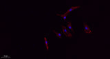
| Validation evidence captions | Immunofluorescence analysis of A549. 1,primary Antibody(red) was diluted at 1:200(4°C overnight). 2, Goat Anti Rabbit IgG (H&L) - Alexa Fluor 594 Secondary antibody was diluted at 1:1000(room temperature, 50min).3, Picture B: DAPI(blue) 10min. |
Key Facts
| Target | CD63 |
| Also known as | CD63; MLA1; TSPAN30; CD63 antigen; Granulophysin; Lysosomal-associated membrane protein 3; LAMP-3; Melanoma-associated antigen ME491; OMA81H; Ocular melanoma-associated antigen; Tetraspanin-30; Tspan-30; CD63 |
| Concentration | 1 mg/ml |
| Working concentration | IF: 1:50-200 Western Blot: 1/500 - 1/2000. IHC-p: 1:100-1:300. ELISA: 1/20000. Not yet tested in other applications. |
| Species reactivity | Human;Rat;Mouse; |
| Observed band | 26,35-65(kD |
| Cellular localization | Cell membrane ; Multi-pass membrane protein . Lysosome membrane ; Multi-pass membrane protein . Late endosome membrane ; Multi-pass membrane protein . Endosome, multivesicular body . Melanosome . Secreted, extracellular exosome . Cell surface . Also found in Weibel-Palade bodies of endothelial cells (PubMed:10793155). Located in platelet dense granules (PubMed:7682577). Detected in a subset of pre-melanosomes. Detected on intralumenal vesicles (ILVs) within multivesicular bodies (PubMed:21962903). . |
| Purity | The antibody was affinity-purified from rabbit antiserum by affinity-chromatography using epitope-specific immunogen. |
| Form / buffer | Liquid in PBS containing 50% glycerol, 0.5% BSA and 0.02% sodium azide. |
| Research area | >>Lysosome;>>Proteoglycans in cancer |
| Host species | Rabbit |
| Clonality | Polyclonal |
| Isotype | IgG |
| Conjugation | Unconjugated |
| Tested applications |
WBIHCIFELISA |
| Dilution range | IF: 1:50-200 Western Blot: 1/500 - 1/2000. IHC-p: 1:100-1:300. ELISA: 1/20000. Not yet tested in other applications. |
| Immunogen | The antiserum was produced against synthesized peptide derived from the Internal region of human CD63. AA range:121-170 |
| Purification | The antibody was affinity-purified from rabbit antiserum by affinity-chromatography using epitope-specific immunogen. |
Reactivity & Application Validation
3 speciess| Species | WB | IHC | IF | ELISA | IP | FCM | CHIP |
|---|---|---|---|---|---|---|---|
| Human | |||||||
| Rat | |||||||
| Mouse |
| Species | Dilution | Notes |
|---|---|---|
| Human | IF: 1:50-200 Western Blot: 1/500 - 1/2000. IHC-p: 1:100-1:300. ELISA: 1/20000. Not yet tested in other applications. | — |
| Rat | — | — |
| Mouse | — | — |
| Species | Dilution | Notes |
|---|
| Species | Dilution | Notes |
|---|
| Species | Dilution | Notes |
|---|
Specifications
Storage & Stability
Compliance & Certifications
Manufactured under ISO 9001:2015 quality management standards.
Not intended for diagnostic or therapeutic use.