Bag-1 rabbit pAb
Bag-1 rabbit pAb from ELK Biotechnology Read more ›

Product Description
Bag-1 rabbit pAb is an ELK Biotechnology antibody for research workflows involving Bag-1.
Background
The oncogene BCL2 is a membrane protein that blocks a step in a pathway leading to apoptosis or programmed cell death. The protein encoded by this gene binds to BCL2 and is referred to as BCL2-associated athanogene. It enhances the anti-apoptotic effects of BCL2 and represents a link between growth factor receptors and anti-apoptotic mechanisms. Multiple protein isoforms are encoded by this mRNA through the use of a non-AUG (CUG) initiation codon, and three alternative downstream AUG initiation codons. A related pseudogene has been defined on chromosome X. [provided by RefSeq, Feb 2010],
Additional antibody information
| Specificity | Bag-1 Polyclonal Antibody detects endogenous levels of Bag-1 protein. |
|---|---|
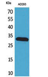
| Validation evidence captions | Western Blot analysis of AD293 cells using Bag-1 Polyclonal Antibody. Secondary antibody(catalog#:RS0002) was diluted at 1:20000 | Western blot analysis of lysate from AD293 cells, using BAG1 Antibody. |
Key Facts
| Target | Bag-1 |
| Also known as | BAG1; HAP; BAG family molecular chaperone regulator 1; BAG-1; Bcl-2-associated athanogene 1 |
| Concentration | 1 mg/ml |
| Working concentration | Western Blot: 1/500 - 1/2000. ELISA: 1/20000. Not yet tested in other applications. |
| Species reactivity | Human;Rat;Mouse; |
| Observed band | 35kD |
| Cellular localization | Isoform 1: Nucleus. Cytoplasm. Isoform 1 localizes predominantly to the nucleus.; Isoform 2: Cytoplasm. Nucleus. Isoform 2 localizes to the cytoplasm and shuttles into the nucleus in response to heat shock.; Isoform 4: Cytoplasm. Nucleus. Isoform 4 localizes predominantly to the cytoplasm. The cellular background in which it is expressed can influence whether it resides primarily in the cytoplasm or is also found in the nucleus. In the presence of BCL2, localizes to intracellular membranes (what appears to be the nuclear envelope and perinuclear membranes) as well as punctate cytosolic structures suggestive of mitochondria. |
| Purity | The antibody was affinity-purified from rabbit antiserum by affinity-chromatography using epitope-specific immunogen. |
| Form / buffer | Liquid in PBS containing 50% glycerol, 0.5% BSA and 0.02% sodium azide. |
| Research area | >>Protein processing in endoplasmic reticulum |
| Host species | Rabbit |
| Clonality | Polyclonal |
| Isotype | IgG |
| Conjugation | Unconjugated |
| Tested applications |
WBELISA |
| Dilution range | Western Blot: 1/500 - 1/2000. ELISA: 1/20000. Not yet tested in other applications. |
| Immunogen | The antiserum was produced against synthesized peptide derived from the Internal region of human BAG1. AA range:41-90 |
| Purification | The antibody was affinity-purified from rabbit antiserum by affinity-chromatography using epitope-specific immunogen. |
Reactivity & Application Validation
3 speciess| Species | WB | IHC | IF | ELISA | IP | FCM | CHIP |
|---|---|---|---|---|---|---|---|
| Human | |||||||
| Rat | |||||||
| Mouse |
| Species | Dilution | Notes |
|---|---|---|
| Human | Western Blot: 1/500 - 1/2000. ELISA: 1/20000. Not yet tested in other applications. | — |
| Rat | — | — |
| Mouse | — | — |
| Species | Dilution | Notes |
|---|
Specifications
Storage & Stability
Compliance & Certifications
Manufactured under ISO 9001:2015 quality management standards.
Not intended for diagnostic or therapeutic use.