Fascin 1 rabbit pAb
Fascin 1 rabbit pAb from ELK Biotechnology Read more ›

Product Description
Fascin 1 rabbit pAb is an ELK Biotechnology antibody for research workflows involving Fascin.
Background
This gene encodes a member of the fascin family of actin-binding proteins. Fascin proteins organize F-actin into parallel bundles, and are required for the formation of actin-based cellular protrusions. The encoded protein plays a critical role in cell migration, motility, adhesion and cellular interactions. Expression of this gene is known to be regulated by several microRNAs, and overexpression of this gene may play a role in the metastasis of multiple types of cancer by increasing cell motility. Expression of this gene is also a marker for Reed-Sternberg cells in Hodgkin's lymphoma. A pseudogene of this gene is located on the long arm of chromosome 15. [provided by RefSeq, Sep 2011],
Additional antibody information
| Specificity | Fascin 1 Polyclonal Antibody detects endogenous levels of Fascin 1 protein. |
|---|---|
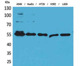
| Validation evidence captions | Western Blot analysis of A549, HuvEc, HT29, K562, L929 cells using Fascin 1 Polyclonal Antibody. Secondary antibody(catalog#:RS0002) was diluted at 1:20000 | Immunohistochemical analysis of paraffin-embedded rat-brain, antibody was diluted at 1:100 | Immunohistochemical analysis of paraffin-embedded mouse-brain, antibody was diluted at 1:100 | Immunohistochemical analysis of paraffin-embedded mouse-brain, antibody was diluted at 1:100 |
Key Facts
| Target | Fascin |
| Also known as | FSCN1; FAN1; HSN; SNL; Fascin; 55 kDa actin-bundling protein; Singed-like protein; p55 |
| Concentration | 1 mg/ml |
| Working concentration | Western Blot: 1/500 - 1/2000. IHC-p: 1:100-300 ELISA: 1/20000. Not yet tested in other applications. |
| Species reactivity | Human;Mouse;Rat |
| Observed band | 55kD |
| Cellular localization | Cytoplasm, cytosol . Cytoplasm, cell cortex . Cytoplasm, cytoskeleton . Cytoplasm, cytoskeleton, stress fiber . Cell projection, filopodium . Cell projection, invadopodium . Cell projection, microvillus . Cell junction . Colocalized with RUFY3 and F-actin at filipodia of the axonal growth cone. Colocalized with DBN1 and F-actin at the transitional domain of the axonal growth cone (By similarity). . |
| Purity | The antibody was affinity-purified from rabbit antiserum by affinity-chromatography using epitope-specific immunogen. |
| Form / buffer | Liquid in PBS containing 50% glycerol, 0.5% BSA and 0.02% sodium azide. |
| Research area | >>MicroRNAs in cancer |
| Host species | Rabbit |
| Clonality | Polyclonal |
| Isotype | IgG |
| Conjugation | Unconjugated |
| Tested applications |
WBIHCIFELISA |
| Dilution range | Western Blot: 1/500 - 1/2000. IHC-p: 1:100-300 ELISA: 1/20000. Not yet tested in other applications. |
| Immunogen | The antiserum was produced against synthesized peptide derived from the Internal region of human FSCN1. AA range:261-310 |
| Purification | The antibody was affinity-purified from rabbit antiserum by affinity-chromatography using epitope-specific immunogen. |
Reactivity & Application Validation
3 speciess| Species | WB | IHC | IF | ELISA | IP | FCM | CHIP |
|---|---|---|---|---|---|---|---|
| Human | |||||||
| Mouse | |||||||
| Rat |
| Species | Dilution | Notes |
|---|---|---|
| Human | Western Blot: 1/500 - 1/2000. IHC-p: 1:100-300 ELISA: 1/20000. Not yet tested in other applications. | — |
| Mouse | — | — |
| Rat | — | — |
| Species | Dilution | Notes |
|---|
| Species | Dilution | Notes |
|---|
| Species | Dilution | Notes |
|---|
Specifications
Storage & Stability
Compliance & Certifications
Manufactured under ISO 9001:2015 quality management standards.
Not intended for diagnostic or therapeutic use.