Stathmin-2 rabbit pAb
Stathmin-2 rabbit pAb from ELK Biotechnology Read more ›

Product Description
Stathmin-2 rabbit pAb is an ELK Biotechnology antibody for research workflows involving Stathmin-2.
Background
This gene encodes a member of the stathmin family of phosphoproteins. Stathmin proteins function in microtubule dynamics and signal transduction. The encoded protein plays a regulatory role in neuronal growth and is also thought to be involved in osteogenesis. Reductions in the expression of this gene have been associated with Down's syndrome and Alzheimer's disease. Alternatively spliced transcript variants have been observed for this gene. A pseudogene of this gene is located on the long arm of chromosome 6. [provided by RefSeq, Nov 2010],
Additional antibody information
| Specificity | Stathmin-2 Polyclonal Antibody detects endogenous levels of Stathmin-2 protein. |
|---|---|
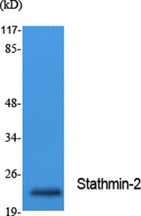
| Validation evidence captions | Western Blot analysis of extracts from Jurkat cells, using Stathmin-2 Polyclonal Antibody. Secondary antibody(catalog#:RS0002) was diluted at 1:20000 | Immunohistochemical analysis of paraffin-embedded rat-brain, antibody was diluted at 1:100 | Immunohistochemical analysis of paraffin-embedded rat-brain, antibody was diluted at 1:100 | Immunohistochemical analysis of paraffin-embedded mouse-brain, antibody was diluted at 1:100 |
Key Facts
| Target | Stathmin-2 |
| Also known as | STMN2; SCG10; SCGN10; Stathmin-2; Superior cervical ganglion-10 protein; Protein SCG10 |
| Concentration | 1 mg/ml |
| Working concentration | Western Blot: 1/500 - 1/2000. IHC-p: 1:100-300 ELISA: 1/5000. Not yet tested in other applications. |
| Species reactivity | Human;Mouse;Rat |
| Observed band | 20kD |
| Cellular localization | Cytoplasm . Cytoplasm, perinuclear region . Cell projection, growth cone. Membrane ; Peripheral membrane protein ; Cytoplasmic side . Cell projection, axon. Golgi apparatus. Endosome . Cell projection, lamellipodium. Associated with punctate structures in the perinuclear cytoplasm, axons, and growth cones of developing neurons. SCG10 exists in both soluble and membrane-bound forms. Colocalized with CIB1 in neurites of developing hippocampal primary neurons (By similarity). Colocalized with CIB1 in the cell body, neuritis and growth cones of neurons. Colocalized with CIB1 to the leading edge of lamellipodia. . |
| Purity | The antibody was affinity-purified from rabbit antiserum by affinity-chromatography using epitope-specific immunogen. |
| Form / buffer | Liquid in PBS containing 50% glycerol, 0.5% BSA and 0.02% sodium azide. |
| Research area | Antibody Research |
| Host species | Rabbit |
| Clonality | Polyclonal |
| Isotype | IgG |
| Conjugation | Unconjugated |
| Tested applications |
WBIHCIFELISA |
| Dilution range | Western Blot: 1/500 - 1/2000. IHC-p: 1:100-300 ELISA: 1/5000. Not yet tested in other applications. |
| Immunogen | Synthesized peptide derived from the Internal region of human Stathmin-2. |
| Purification | The antibody was affinity-purified from rabbit antiserum by affinity-chromatography using epitope-specific immunogen. |
Reactivity & Application Validation
3 speciess| Species | WB | IHC | IF | ELISA | IP | FCM | CHIP |
|---|---|---|---|---|---|---|---|
| Human | |||||||
| Mouse | |||||||
| Rat |
| Species | Dilution | Notes |
|---|---|---|
| Human | Western Blot: 1/500 - 1/2000. IHC-p: 1:100-300 ELISA: 1/5000. Not yet tested in other applications. | — |
| Mouse | — | — |
| Rat | — | — |
| Species | Dilution | Notes |
|---|
| Species | Dilution | Notes |
|---|
| Species | Dilution | Notes |
|---|
Specifications
Storage & Stability
Compliance & Certifications
Manufactured under ISO 9001:2015 quality management standards.
Not intended for diagnostic or therapeutic use.