Myosin VI rabbit pAb
Myosin VI rabbit pAb from ELK Biotechnology Read more ›

Product Description
Myosin VI rabbit pAb is an ELK Biotechnology antibody for research workflows involving Myosin VI.
Background
myosin VI(MYO6) Homo sapiens This gene encodes a reverse-direction motor protein that moves toward the minus end of actin filaments and plays a role in intracellular vesicle and organelle transport. The protein consists of a motor domain containing an ATP- and an actin-binding site and a globular tail which interacts with other proteins. This protein maintains the structural integrity of inner ear hair cells and mutations in this gene cause non-syndromic autosomal dominant and recessive hearing loss. Alternative splicing results in multiple transcript variants encoding distinct isoforms. [provided by RefSeq, Jul 2014],
Additional antibody information
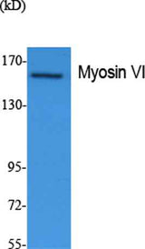
| Specificity | Myosin VI Polyclonal Antibody detects endogenous levels of Myosin VI protein. |
|---|---|
| Validation evidence captions | Western Blot analysis of extracts from Jurkat cells, using Myosin VI Polyclonal Antibody. Secondary antibody(catalog#:RS0002) was diluted at 1:20000 | Immunohistochemical analysis of paraffin-embedded rat-brain, antibody was diluted at 1:100 | Immunohistochemical analysis of paraffin-embedded rat-brain, antibody was diluted at 1:100 | Immunohistochemical analysis of paraffin-embedded rat-brain, antibody was diluted at 1:100 |
Key Facts
| Target | Myosin VI |
| Also known as | MYO6; KIAA0389; Unconventional myosin-VI; Unconventional myosin-6 |
| Concentration | 1 mg/ml |
| Working concentration | Western Blot: 1/500 - 1/2000. IHC-p: 1:100-300 ELISA: 1/5000. Not yet tested in other applications. |
| Species reactivity | Human;Mouse;Rat |
| Observed band | 149kD |
| Cellular localization | Golgi apparatus, trans-Golgi network membrane ; Peripheral membrane protein . Golgi apparatus . Nucleus . Cytoplasm, perinuclear region . Membrane, clathrin-coated pit . Cytoplasmic vesicle, clathrin-coated vesicle . Cell projection, filopodium . Cell projection, ruffle membrane . Cell projection, microvillus . Cytoplasm, cytosol . Also present in endocyctic vesicles (PubMed:16507995). Translocates from membrane ruffles, endocytic vesicles and cytoplasm to Golgi apparatus, perinuclear membrane and nucleus through induction by p53 and p53-induced DNA damage (PubMed:16507995). Recruited into membrane ruffles from cell surface by EGF-stimulation (PubMed:9852149). Colocalizes with DAB2 in clathrin-coated pits/vesicles (PubMed:11967127). Colocalizes with OPTN at the Golgi complex and in vesicul |
| Purity | The antibody was affinity-purified from rabbit antiserum by affinity-chromatography using epitope-specific immunogen. |
| Form / buffer | Liquid in PBS containing 50% glycerol, 0.5% BSA and 0.02% sodium azide. |
| Research area | >>Pathogenic Escherichia coli infection;>>Salmonella infection |
| Host species | Rabbit |
| Clonality | Polyclonal |
| Isotype | IgG |
| Conjugation | Unconjugated |
| Tested applications |
WBIHCIFELISA |
| Dilution range | Western Blot: 1/500 - 1/2000. IHC-p: 1:100-300 ELISA: 1/5000. Not yet tested in other applications. |
| Immunogen | Synthesized peptide derived from Myosin VI . at AA range: 40-120 |
| Purification | The antibody was affinity-purified from rabbit antiserum by affinity-chromatography using epitope-specific immunogen. |
Reactivity & Application Validation
3 speciess| Species | WB | IHC | IF | ELISA | IP | FCM | CHIP |
|---|---|---|---|---|---|---|---|
| Human | |||||||
| Mouse | |||||||
| Rat |
| Species | Dilution | Notes |
|---|---|---|
| Human | Western Blot: 1/500 - 1/2000. IHC-p: 1:100-300 ELISA: 1/5000. Not yet tested in other applications. | — |
| Mouse | — | — |
| Rat | — | — |
| Species | Dilution | Notes |
|---|
| Species | Dilution | Notes |
|---|
| Species | Dilution | Notes |
|---|
Specifications
Storage & Stability
Compliance & Certifications
Manufactured under ISO 9001:2015 quality management standards.
Not intended for diagnostic or therapeutic use.