V-ATPase D1 rabbit pAb
V-ATPase D1 rabbit pAb from ELK Biotechnology Read more ›

Product Description
V-ATPase D1 rabbit pAb is an ELK Biotechnology antibody for research workflows involving V-ATPase D1.
Background
This gene encodes a component of vacuolar ATPase (V-ATPase), a multisubunit enzyme that mediates acidification of eukaryotic intracellular organelles. V-ATPase dependent organelle acidification is necessary for such intracellular processes as protein sorting, zymogen activation, receptor-mediated endocytosis, and synaptic vesicle proton gradient generation. V-ATPase is composed of a cytosolic V1 domain and a transmembrane V0 domain. The V1 domain consists of three A and three B subunits, two G subunits plus the C, D, E, F, and H subunits. The V1 domain contains the ATP catalytic site. The V0 domain consists of five different subunits: a, c, c', c'', and d. Additional isoforms of many of the V1 and V0 subunit proteins are encoded by multiple genes or alternatively spliced transcript variants. This encoded protein is known as the D subunit and is found ubiquitously. [pro
Additional antibody information
| Specificity | V-ATPase D1 Polyclonal Antibody detects endogenous levels of V-ATPase D1 protein. |
|---|---|
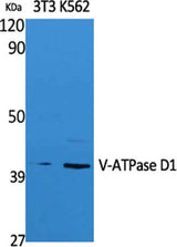
| Validation evidence captions | Western Blot analysis of extracts from NIH-3T3, K562 cells, using V-ATPase D1 Polyclonal Antibody. Secondary antibody(catalog#:RS0002) was diluted at 1:20000 | Western blot analysis of lysates from HeLa cells, using V-ATPase D1 antibody. |
Key Facts
| Target | V-ATPase D1 |
| Also known as | ATP6V0D1; ATP6D; VPATPD; V-type proton ATPase subunit d 1; V-ATPase subunit d 1; 32 kDa accessory protein; V-ATPase 40 kDa accessory protein; V-ATPase AC39 subunit; p39; Vacuolar proton pump subunit d 1 |
| Concentration | 1 mg/ml |
| Working concentration | Western Blot: 1/500 - 1/2000. ELISA: 1/20000. Not yet tested in other applications. |
| Species reactivity | Human;Mouse;Rat |
| Observed band | 40kD |
| Cellular localization | Membrane ; Peripheral membrane protein ; Cytoplasmic side . Lysosome membrane ; Peripheral membrane protein . Cytoplasmic vesicle, clathrin-coated vesicle membrane ; Peripheral membrane protein . Localizes to centrosome and the base of the cilium. . |
| Purity | The antibody was affinity-purified from rabbit antiserum by affinity-chromatography using epitope-specific immunogen. |
| Form / buffer | Liquid in PBS containing 50% glycerol, 0.5% BSA and 0.02% sodium azide. |
| Research area | >>Oxidative phosphorylation;>>Metabolic pathways;>>Lysosome;>>Phagosome;>>Synaptic vesicle cycle;>>Collecting duct acid secretion;>>Vibrio cholerae infection;>>Epithelial cell signaling in Helicobacter pylori infection;>>Tuberculosis;>>Human papillomavirus infection;>>Viral carcinogenesis;>>Rheumatoid arthritis |
| Host species | Rabbit |
| Clonality | Polyclonal |
| Isotype | IgG |
| Conjugation | Unconjugated |
| Tested applications |
WBELISA |
| Dilution range | Western Blot: 1/500 - 1/2000. ELISA: 1/20000. Not yet tested in other applications. |
| Immunogen | The antiserum was produced against synthesized peptide derived from human V-ATPase D1. AA range:221-270 |
| Purification | The antibody was affinity-purified from rabbit antiserum by affinity-chromatography using epitope-specific immunogen. |
Reactivity & Application Validation
3 speciess| Species | WB | IHC | IF | ELISA | IP | FCM | CHIP |
|---|---|---|---|---|---|---|---|
| Human | |||||||
| Mouse | |||||||
| Rat |
| Species | Dilution | Notes |
|---|---|---|
| Human | Western Blot: 1/500 - 1/2000. ELISA: 1/20000. Not yet tested in other applications. | — |
| Mouse | — | — |
| Rat | — | — |
| Species | Dilution | Notes |
|---|
Specifications
Storage & Stability
Compliance & Certifications
Manufactured under ISO 9001:2015 quality management standards.
Not intended for diagnostic or therapeutic use.