RXRγ rabbit pAb
RXRγ rabbit pAb from ELK Biotechnology Read more ›

Product Description
RXRγ rabbit pAb is an ELK Biotechnology antibody for research workflows involving RXRγ.
Background
retinoid X receptor gamma(RXRG) Homo sapiens This gene encodes a member of the retinoid X receptor (RXR) family of nuclear receptors which are involved in mediating the antiproliferative effects of retinoic acid (RA). This receptor forms dimers with the retinoic acid, thyroid hormone, and vitamin D receptors, increasing both DNA binding and transcriptional function on their respective response elements. This gene is expressed at significantly lower levels in non-small cell lung cancer cells. Alternatively spliced transcript variants have been described. [provided by RefSeq, Jun 2010],
Additional antibody information
| Specificity | RXRγ Polyclonal Antibody detects endogenous levels of RXRγ protein. |
|---|---|
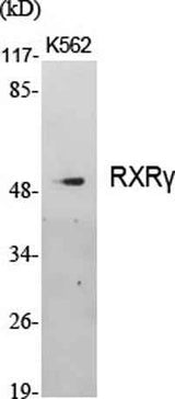
| Validation evidence captions | Western Blot analysis of various cells using RXRγ Polyclonal Antibody cells nucleus extracted by Minute TM Cytoplasmic and Nuclear Fractionation kit (SC-003,Inventbiotech,MN,USA). | Western Blot analysis of HepG2 cells using RXRγ Polyclonal Antibody cells nucleus extracted by Minute TM Cytoplasmic and Nuclear Fractionation kit (SC-003,Inventbiotech,MN,USA). | Immunohistochemical analysis of paraffin-embedded Human lung cancer. Antibody was diluted at 1:100(4° overnight). High-pressure and temperature Tris-EDTA,pH8.0 was used for antigen retrieval. | Immunofluorescence analysis of HUVEC cells, using Retinoid X Receptor gamma Antibody. The picture on the right is blocked with the synthesized peptide. |
Key Facts
| Target | RXRγ |
| Also known as | RXRG; NR2B3; Retinoic acid receptor RXR-gamma; Nuclear receptor subfamily 2 group B member 3; Retinoid X receptor gamma |
| Concentration | 1 mg/ml |
| Working concentration | Western Blot: 1/500 - 1/2000. Immunohistochemistry: 1/100 - 1/300. Immunofluorescence: 1/200 - 1/1000. ELISA: 1/10000. Not yet tested in other applications. |
| Species reactivity | Human;Mouse |
| Observed band | 50kD |
| Cellular localization | Nucleus . Cytoplasm . |
| Purity | The antibody was affinity-purified from rabbit antiserum by affinity-chromatography using epitope-specific immunogen. |
| Form / buffer | Liquid in PBS containing 50% glycerol, 0.5% BSA and 0.02% sodium azide. |
| Research area | >>PPAR signaling pathway;>>Th17 cell differentiation;>>Thyroid hormone signaling pathway;>>Adipocytokine signaling pathway;>>Parathyroid hormone synthesis, secretion and action;>>Pathways in cancer;>>Transcriptional misregulation in cancer;>>Chemical carcinogenesis - receptor activation;>>Thyroid cancer;>>Small cell lung cancer;>>Non-small cell lung cancer;>>Gastric cancer;>>Lipid and atherosclerosis |
| Host species | Rabbit |
| Clonality | Polyclonal |
| Isotype | IgG |
| Conjugation | Unconjugated |
| Tested applications |
WBIHCIFELISA |
| Dilution range | Western Blot: 1/500 - 1/2000. Immunohistochemistry: 1/100 - 1/300. Immunofluorescence: 1/200 - 1/1000. ELISA: 1/10000. Not yet tested in other applications. |
| Immunogen | The antiserum was produced against synthesized peptide derived from human Retinoid X Receptor gamma. AA range:171-220 |
| Purification | The antibody was affinity-purified from rabbit antiserum by affinity-chromatography using epitope-specific immunogen. |
Reactivity & Application Validation
2 speciess| Species | WB | IHC | IF | ELISA | IP | FCM | CHIP |
|---|---|---|---|---|---|---|---|
| Human | |||||||
| Mouse |
| Species | Dilution | Notes |
|---|---|---|
| Human | Western Blot: 1/500 - 1/2000. Immunohistochemistry: 1/100 - 1/300. Immunofluorescence: 1/200 - 1/1000. ELISA: 1/10000. Not yet tested in other applications. | — |
| Mouse | — | — |
| Species | Dilution | Notes |
|---|
| Species | Dilution | Notes |
|---|
| Species | Dilution | Notes |
|---|
Specifications
Storage & Stability
Compliance & Certifications
Manufactured under ISO 9001:2015 quality management standards.
Not intended for diagnostic or therapeutic use.