Ret rabbit pAb
Ret rabbit pAb from ELK Biotechnology Read more ›

Product Description
Ret rabbit pAb is an ELK Biotechnology antibody for research workflows involving Ret.
Background
ret proto-oncogene(RET) Homo sapiens This gene, a member of the cadherin superfamily, encodes one of the receptor tyrosine kinases, which are cell-surface molecules that transduce signals for cell growth and differentiation. This gene plays a crucial role in neural crest development, and it can undergo oncogenic activation in vivo and in vitro by cytogenetic rearrangement. Mutations in this gene are associated with the disorders multiple endocrine neoplasia, type IIA, multiple endocrine neoplasia, type IIB, Hirschsprung disease, and medullary thyroid carcinoma. Two transcript variants encoding different isoforms have been found for this gene. Additional transcript variants have been described but their biological validity has not been confirmed. [provided by RefSeq, Jul 2008],
Additional antibody information
| Specificity | Ret Polyclonal Antibody detects endogenous levels of Ret protein. |
|---|---|
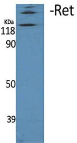
| Validation evidence captions | Western Blot analysis of various cells using Ret Polyclonal Antibody | Immunofluorescence analysis of HUVEC cells, using Ret Antibody. The picture on the right is blocked with the synthesized peptide. | Immunohistochemistry analysis of paraffin-embedded human brain, using Ret Antibody. The picture on the right is blocked with the synthesized peptide. | Western blot analysis of lysates from Jurkat cells, using Ret Antibody. The lane on the right is blocked with the synthesized peptide. |
Key Facts
| Target | Ret |
| Also known as | RET; CDHF12; CDHR16; PTC; RET51; Proto-oncogene tyrosine-protein kinase receptor Ret; Cadherin family member 12; Proto-oncogene c-Ret |
| Concentration | 1 mg/ml |
| Working concentration | Western Blot: 1/500 - 1/2000. Immunohistochemistry: 1/100 - 1/300. Immunofluorescence: 1/200 - 1/1000. ELISA: 1/20000. Not yet tested in other applications. |
| Species reactivity | Human;Mouse;Rat |
| Observed band | 170kD |
| Cellular localization | Cell membrane ; Single-pass type I membrane protein . Endosome membrane ; Single-pass type I membrane protein . Predominantly located on the plasma membrane. In the presence of SORL1 and GFRA1, directed to endosomes. . |
| Purity | The antibody was affinity-purified from rabbit antiserum by affinity-chromatography using epitope-specific immunogen. |
| Form / buffer | Liquid in PBS containing 50% glycerol, 0.5% BSA and 0.02% sodium azide. |
| Research area | >>Calcium signaling pathway;>>Pathways in cancer;>>Thyroid cancer;>>Non-small cell lung cancer;>>Central carbon metabolism in cancer |
| Host species | Rabbit |
| Clonality | Polyclonal |
| Isotype | IgG |
| Conjugation | Unconjugated |
| Tested applications |
WBIHCIFELISA |
| Dilution range | Western Blot: 1/500 - 1/2000. Immunohistochemistry: 1/100 - 1/300. Immunofluorescence: 1/200 - 1/1000. ELISA: 1/20000. Not yet tested in other applications. |
| Immunogen | The antiserum was produced against synthesized peptide derived from human RET. AA range:881-930 |
| Purification | The antibody was affinity-purified from rabbit antiserum by affinity-chromatography using epitope-specific immunogen. |
Reactivity & Application Validation
3 speciess| Species | WB | IHC | IF | ELISA | IP | FCM | CHIP |
|---|---|---|---|---|---|---|---|
| Human | |||||||
| Mouse | |||||||
| Rat |
| Species | Dilution | Notes |
|---|---|---|
| Human | Western Blot: 1/500 - 1/2000. Immunohistochemistry: 1/100 - 1/300. Immunofluorescence: 1/200 - 1/1000. ELISA: 1/20000. Not yet tested in other applications. | — |
| Mouse | — | — |
| Rat | — | — |
| Species | Dilution | Notes |
|---|
| Species | Dilution | Notes |
|---|
| Species | Dilution | Notes |
|---|
Specifications
Storage & Stability
Compliance & Certifications
Manufactured under ISO 9001:2015 quality management standards.
Not intended for diagnostic or therapeutic use.