PIPK I γ rabbit pAb
PIPK I γ rabbit pAb from ELK Biotechnology Read more ›

Product Description
PIPK I γ rabbit pAb is an ELK Biotechnology antibody for research workflows involving PIPK I γ.
Background
phosphatidylinositol-4-phosphate 5-kinase type 1 gamma(PIP5K1C) Homo sapiens This locus encodes a type I phosphatidylinositol 4-phosphate 5-kinase. The encoded protein catalyzes phosphorylation of phosphatidylinositol 4-phosphate, producing phosphatidylinositol 4,5-bisphosphate. This enzyme is found at synapses and has been found to play roles in endocytosis and cell migration. Mutations at this locus have been associated with lethal congenital contractural syndrome. Alternatively spliced transcript variants encoding different isoforms have been described.[provided by RefSeq, Sep 2010],
Additional antibody information
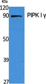
| Specificity | PIPK I γ Polyclonal Antibody detects endogenous levels of PIPK I γ protein. |
|---|---|
| Validation evidence captions | Western Blot analysis of various cells using PIPK I γ Polyclonal Antibody | Western Blot analysis of A549 cells using PIPK I γ Polyclonal Antibody | Western blot analysis of lysates from A549 cells, using PIP5K1C Antibody. The lane on the right is blocked with the synthesized peptide. | Western blot analysis of the lysates from HepG2 cells using PIP5K1C antibody. |
Key Facts
| Target | PIPK I γ |
| Also known as | PIP5K1C; KIAA0589; Phosphatidylinositol 4-phosphate 5-kinase type-1 gamma; PIP5K1-gamma; PtdIns(4)P-5-kinase 1 gamma; Phosphatidylinositol 4-phosphate 5-kinase type I gamma; PIP5KIgamma |
| Concentration | 1 mg/ml |
| Working concentration | Western Blot: 1/500 - 1/2000. ELISA: 1/20000. Not yet tested in other applications. |
| Species reactivity | Human;Rat;Mouse; |
| Observed band | 80kD |
| Cellular localization | Cell membrane; Peripheral membrane protein; Cytoplasmic side . Endomembrane system . Cytoplasm . Cell junction, focal adhesion . Cell junction, adherens junction . Cell projection, ruffle membrane . Cell projection, phagocytic cup . Cell projection, uropodium . Detected in plasma membrane invaginations. Isoform 3 is detected in intracellular vesicle-like structures.; Isoform 2: Cytoplasm. Nucleus. |
| Purity | The antibody was affinity-purified from rabbit antiserum by affinity-chromatography using epitope-specific immunogen. |
| Form / buffer | Liquid in PBS containing 50% glycerol, 0.5% BSA and 0.02% sodium azide. |
| Research area | >>Inositol phosphate metabolism;>>Metabolic pathways;>>Phosphatidylinositol signaling system;>>Phospholipase D signaling pathway;>>Endocytosis;>>Focal adhesion;>>Fc gamma R-mediated phagocytosis;>>Regulation of actin cytoskeleton;>>Yersinia infection;>>Choline metabolism in cancer |
| Host species | Rabbit |
| Clonality | Polyclonal |
| Isotype | IgG |
| Conjugation | Unconjugated |
| Tested applications |
WBELISA |
| Dilution range | Western Blot: 1/500 - 1/2000. ELISA: 1/20000. Not yet tested in other applications. |
| Immunogen | The antiserum was produced against synthesized peptide derived from human PIP5K1C. AA range:305-354 |
| Purification | The antibody was affinity-purified from rabbit antiserum by affinity-chromatography using epitope-specific immunogen. |
Reactivity & Application Validation
3 speciess| Species | WB | IHC | IF | ELISA | IP | FCM | CHIP |
|---|---|---|---|---|---|---|---|
| Human | |||||||
| Rat | |||||||
| Mouse |
| Species | Dilution | Notes |
|---|---|---|
| Human | Western Blot: 1/500 - 1/2000. ELISA: 1/20000. Not yet tested in other applications. | — |
| Rat | — | — |
| Mouse | — | — |
| Species | Dilution | Notes |
|---|
Specifications
Storage & Stability
Compliance & Certifications
Manufactured under ISO 9001:2015 quality management standards.
Not intended for diagnostic or therapeutic use.