PARK7 rabbit pAb
PARK7 rabbit pAb from ELK Biotechnology Read more ›

Product Description
PARK7 rabbit pAb is an ELK Biotechnology antibody for research workflows involving PARK7.
Background
The product of this gene belongs to the peptidase C56 family of proteins. It acts as a positive regulator of androgen receptor-dependent transcription. It may also function as a redox-sensitive chaperone, as a sensor for oxidative stress, and it apparently protects neurons against oxidative stress and cell death. Defects in this gene are the cause of autosomal recessive early-onset Parkinson disease 7. Two transcript variants encoding the same protein have been identified for this gene. [provided by RefSeq, Jul 2008],
Additional antibody information
| Specificity | PARK7 Polyclonal Antibody detects endogenous levels of PARK7 protein. |
|---|---|
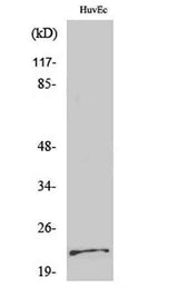
| Validation evidence captions | Western Blot analysis of various cells using PARK7 Polyclonal Antibody | Immunofluorescence analysis of HeLa cells, using DJ-1 Antibody. The picture on the right is blocked with the synthesized peptide. | Immunohistochemistry analysis of paraffin-embedded human lung carcinoma tissue, using DJ-1 Antibody. The picture on the right is blocked with the synthesized peptide. | Western blot analysis of lysates from HUVEC cells, using DJ-1 Antibody. The lane on the right is blocked with the synthesized peptide. |
Key Facts
| Target | PARK7 |
| Also known as | PARK7; Protein DJ-1; Oncogene DJ1; Parkinson disease protein 7 |
| Concentration | 1 mg/ml |
| Working concentration | Western Blot: 1/500 - 1/2000. Immunohistochemistry: 1/100 - 1/300. Immunofluorescence: 1/200 - 1/1000. ELISA: 1/10000. Not yet tested in other applications. |
| Species reactivity | Human;Mouse |
| Observed band | 22kD |
| Cellular localization | Cell membrane ; Lipid-anchor . Cytoplasm . Nucleus . Membrane raft . Mitochondrion . Endoplasmic reticulum . Under normal conditions, located predominantly in the cytoplasm and, to a lesser extent, in the nucleus and mitochondrion. Translocates to the mitochondrion and subsequently to the nucleus in response to oxidative stress and exerts an increased cytoprotective effect against oxidative damage (PubMed:18711745). Detected in tau inclusions in brains from neurodegenerative disease patients (PubMed:14705119). Membrane raft localization in astrocytes and neuronal cells requires palmitoylation. . |
| Purity | The antibody was affinity-purified from rabbit antiserum by affinity-chromatography using epitope-specific immunogen. |
| Form / buffer | Liquid in PBS containing 50% glycerol, 0.5% BSA and 0.02% sodium azide. |
| Research area | >>Parkinson disease;>>Pathways of neurodegeneration - multiple diseases |
| Host species | Rabbit |
| Clonality | Polyclonal |
| Isotype | IgG |
| Conjugation | Unconjugated |
| Tested applications |
WBIHCIFELISA |
| Dilution range | Western Blot: 1/500 - 1/2000. Immunohistochemistry: 1/100 - 1/300. Immunofluorescence: 1/200 - 1/1000. ELISA: 1/10000. Not yet tested in other applications. |
| Immunogen | The antiserum was produced against synthesized peptide derived from human DJ-1. AA range:21-70 |
| Purification | The antibody was affinity-purified from rabbit antiserum by affinity-chromatography using epitope-specific immunogen. |
Reactivity & Application Validation
2 speciess| Species | WB | IHC | IF | ELISA | IP | FCM | CHIP |
|---|---|---|---|---|---|---|---|
| Human | |||||||
| Mouse |
| Species | Dilution | Notes |
|---|---|---|
| Human | Western Blot: 1/500 - 1/2000. Immunohistochemistry: 1/100 - 1/300. Immunofluorescence: 1/200 - 1/1000. ELISA: 1/10000. Not yet tested in other applications. | — |
| Mouse | — | — |
| Species | Dilution | Notes |
|---|
| Species | Dilution | Notes |
|---|
| Species | Dilution | Notes |
|---|
Specifications
Storage & Stability
Compliance & Certifications
Manufactured under ISO 9001:2015 quality management standards.
Not intended for diagnostic or therapeutic use.