PAKα rabbit pAb
PAKα rabbit pAb from ELK Biotechnology Read more ›

Product Description
PAKα rabbit pAb is an ELK Biotechnology antibody for research workflows involving PAK1.
Background
This gene encodes a family member of serine/threonine p21-activating kinases, known as PAK proteins. These proteins are critical effectors that link RhoGTPases to cytoskeleton reorganization and nuclear signaling, and they serve as targets for the small GTP binding proteins Cdc42 and Rac. This specific family member regulates cell motility and morphology. Alternatively spliced transcript variants encoding different isoforms have been found for this gene. [provided by RefSeq, Apr 2010],
Additional antibody information
| Specificity | PAKα Polyclonal Antibody detects endogenous levels of PAKα protein. |
|---|---|
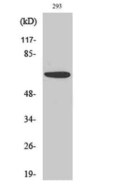
| Validation evidence captions | Western Blot analysis of various cells using PAKα Polyclonal Antibody diluted at 1:500 | Immunofluorescence analysis of HeLa cells, using PAK1 Antibody. The picture on the right is blocked with the synthesized peptide. | Immunohistochemistry analysis of paraffin-embedded human colon carcinoma tissue, using PAK1 Antibody. The picture on the right is blocked with the synthesized peptide. | Western blot analysis of lysates from 293 cells, treated with Etoposide 25uM 60', using PAK1 Antibody. The lane on the right is blocked with the synthesized peptide. |
Key Facts
| Target | PAK1 |
| Also known as | PAK1; Serine/threonine-protein kinase PAK 1; Alpha-PAK; p21-activated kinase 1; PAK-1; p65-PAK |
| Concentration | 1 mg/ml |
| Working concentration | Western Blot: 1/500 - 1/2000. Immunohistochemistry: 1/100 - 1/300. Immunofluorescence: 1/200 - 1/1000. ELISA: 1/5000. Not yet tested in other applications. |
| Species reactivity | Human;Mouse;Rat |
| Observed band | 60kD |
| Cellular localization | Cytoplasm . Cell junction, focal adhesion . Cell projection, lamellipodium . Cell membrane . Cell projection, ruffle membrane . Cell projection, invadopodium . Nucleus, nucleoplasm . Chromosome . Cytoplasm, cytoskeleton, microtubule organizing center, centrosome . Colocalizes with RUFY3, F-actin and other core migration components in invadopodia at the cell periphery (PubMed:25766321). Recruited to the cell membrane by interaction with CDC42 and RAC1. Recruited to focal adhesions upon activation. Colocalized with CIB1 within membrane ruffles during cell spreading upon readhesion to fibronectin. Upon DNA damage, translocates to the nucleoplasm when phosphorylated at Thr-212 where is co-recruited with MORC2 on damaged chromatin (PubMed:23260667). Localization to the centrosome does not depen |
| Purity | The antibody was affinity-purified from rabbit antiserum by affinity-chromatography using epitope-specific immunogen. |
| Form / buffer | Liquid in PBS containing 50% glycerol, 0.5% BSA and 0.02% sodium azide. |
| Research area | >>MAPK signaling pathway;>>ErbB signaling pathway;>>Ras signaling pathway;>>cAMP signaling pathway;>>Chemokine signaling pathway;>>Axon guidance;>>Hippo signaling pathway - multiple species;>>Focal adhesion;>>C-type lectin receptor signaling pathway;>>Natural killer cell mediated cytotoxicity;>>T cell receptor signaling pathway;>>Fc gamma R-mediated phagocytosis;>>Regulation of actin cytoskeleton;>>Epithelial cell signaling in Helicobacter pylori infection;>>Pathogenic Escherichia coli infection;>>Salmonella infection;>>Human immunodeficiency virus 1 infection;>>Proteoglycans in cancer;>>Renal cell carcinoma |
| Host species | Rabbit |
| Clonality | Polyclonal |
| Isotype | IgG |
| Conjugation | Unconjugated |
| Tested applications |
WBIHCIFELISA |
| Dilution range | Western Blot: 1/500 - 1/2000. Immunohistochemistry: 1/100 - 1/300. Immunofluorescence: 1/200 - 1/1000. ELISA: 1/5000. Not yet tested in other applications. |
| Immunogen | The antiserum was produced against synthesized peptide derived from human PAK1. AA range:178-227 |
| Purification | The antibody was affinity-purified from rabbit antiserum by affinity-chromatography using epitope-specific immunogen. |
Reactivity & Application Validation
3 speciess| Species | WB | IHC | IF | ELISA | IP | FCM | CHIP |
|---|---|---|---|---|---|---|---|
| Human | |||||||
| Mouse | |||||||
| Rat |
| Species | Dilution | Notes |
|---|---|---|
| Human | Western Blot: 1/500 - 1/2000. Immunohistochemistry: 1/100 - 1/300. Immunofluorescence: 1/200 - 1/1000. ELISA: 1/5000. Not yet tested in other applications. | — |
| Mouse | — | — |
| Rat | — | — |
| Species | Dilution | Notes |
|---|
| Species | Dilution | Notes |
|---|
| Species | Dilution | Notes |
|---|
Specifications
Storage & Stability
Compliance & Certifications
Manufactured under ISO 9001:2015 quality management standards.
Not intended for diagnostic or therapeutic use.