Gα t1 rabbit pAb
Gα t1 rabbit pAb from ELK Biotechnology Read more ›

Product Description
Gα t1 rabbit pAb is an ELK Biotechnology antibody for research workflows involving Gα t1.
Background
Transducin is a 3-subunit guanine nucleotide-binding protein (G protein) which stimulates the coupling of rhodopsin and cGMP-phoshodiesterase during visual impulses. The transducin alpha subunits in rods and cones are encoded by separate genes. This gene encodes the alpha subunit in rods. This gene is also expressed in other cells, and has been implicated in bitter taste transduction in rat taste cells. Mutations in this gene result in autosomal dominant congenital stationary night blindness. Multiple alternatively spliced variants, encoding the same protein, have been identified. [provided by RefSeq, Feb 2009],
Additional antibody information
| Specificity | Gα t1 Polyclonal Antibody detects endogenous levels of Gα t1 protein. |
|---|---|
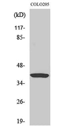
| Validation evidence captions | Western Blot analysis of various cells using Gα t1 Polyclonal Antibody diluted at 1:2000 | Western blot analysis of lysates from COLO cells, using GNAT1 Antibody. The lane on the right is blocked with the synthesized peptide. |
Key Facts
| Target | Gα t1 |
| Also known as | GNAT1; GNATR; Guanine nucleotide-binding protein G(t) subunit alpha-1; Transducin alpha-1 chain |
| Concentration | 1 mg/ml |
| Working concentration | Western Blot: 1/500 - 1/2000. ELISA: 1/40000. Not yet tested in other applications. |
| Species reactivity | Human;Mouse;Rat |
| Observed band | 36kD |
| Cellular localization | Cell projection, cilium, photoreceptor outer segment . Membrane ; Peripheral membrane protein . Photoreceptor inner segment . Localizes mainly in the outer segment in the dark-adapted state, whereas is translocated to the inner part of the photoreceptors in the light-adapted state. During dark-adapted conditions, in the presence of UNC119 mislocalizes from the outer segment to the inner part of rod photoreceptors which leads to decreased photoreceptor damage caused by light. . |
| Purity | The antibody was affinity-purified from rabbit antiserum by affinity-chromatography using epitope-specific immunogen. |
| Form / buffer | Liquid in PBS containing 50% glycerol, 0.5% BSA and 0.02% sodium azide. |
| Research area | >>Phototransduction |
| Host species | Rabbit |
| Clonality | Polyclonal |
| Isotype | IgG |
| Conjugation | Unconjugated |
| Tested applications |
WBELISA |
| Dilution range | Western Blot: 1/500 - 1/2000. ELISA: 1/40000. Not yet tested in other applications. |
| Immunogen | The antiserum was produced against synthesized peptide derived from human GNAT1. AA range:71-120 |
| Purification | The antibody was affinity-purified from rabbit antiserum by affinity-chromatography using epitope-specific immunogen. |
Reactivity & Application Validation
3 speciess| Species | WB | IHC | IF | ELISA | IP | FCM | CHIP |
|---|---|---|---|---|---|---|---|
| Human | |||||||
| Mouse | |||||||
| Rat |
| Species | Dilution | Notes |
|---|---|---|
| Human | Western Blot: 1/500 - 1/2000. ELISA: 1/40000. Not yet tested in other applications. | — |
| Mouse | — | — |
| Rat | — | — |
| Species | Dilution | Notes |
|---|
Specifications
Storage & Stability
Compliance & Certifications
Manufactured under ISO 9001:2015 quality management standards.
Not intended for diagnostic or therapeutic use.