FAS-L rabbit pAb
FAS-L rabbit pAb from ELK Biotechnology Read more ›

Product Description
FAS-L rabbit pAb is an ELK Biotechnology antibody for research workflows involving FAS-L.
Background
This gene is a member of the tumor necrosis factor superfamily. The primary function of the encoded transmembrane protein is the induction of apoptosis triggered by binding to FAS. The FAS/FASLG signaling pathway is essential for immune system regulation, including activation-induced cell death (AICD) of T cells and cytotoxic T lymphocyte induced cell death. It has also been implicated in the progression of several cancers. Defects in this gene may be related to some cases of systemic lupus erythematosus (SLE). Alternatively spliced transcript variants have been described. [provided by RefSeq, Nov 2014],
Additional antibody information
| Specificity | FAS-L Polyclonal Antibody detects endogenous levels of FAS-L protein. |
|---|---|
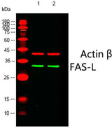
| Validation evidence captions | Western blot analysis of lysates from 1)HepG2, 2)293 cells, (Green) primary antibody was diluted at 1:1000, 4°over night, Dylight 800 secondary antibody(Immunoway:RS23920)was diluted at 1:10000, 37° 1hour. (Red) Actin β Monoclonal Antibody(5G3) (Immunoway:YM3028) antibody was diluted at 1:5000 as loading control, 4° over night,Dylight 680 secondary antibody(Immunoway:RS23710)was diluted at 1:10000, 37° 1hour. | Immunofluorescence analysis of rat-lung tissue. 1,FAS-L Polyclonal Antibody(red) was diluted at 1:200(4°C,overnight). 2, Cy3 labled Secondary antibody was diluted at 1:300(room temperature, 50min).3, Picture B: DAPI(blue) 10min. Picture A:Target. Picture B: DAPI. Picture C: merge of A+B | Immunofluorescence analysis of rat-lung tissue. 1,FAS-L Polyclonal Antibody(red) was diluted at 1:200(4°C,overnight). 2, Cy3 labled Secondary antibody was diluted at 1:300(room temperature, 50min).3, Picture B: DAPI(blue) 10min. Picture A:Target. Picture B: DAPI. Picture C: merge of A+B | Immunofluorescence analysis of rat-kidney tissue. 1,FAS-L Polyclonal Antibody(red) was diluted at 1:200(4°C,overnight). 2, Cy3 labled Secondary antibody was diluted at 1:300(room temperature, 50min).3, Picture B: DAPI(blue) 10min. Picture A:Target. Picture B: DAPI. Picture C: merge of A+B |
Key Facts
| Target | FAS-L |
| Also known as | FASLG; APT1LG1; CD95L; FASL; TNFSF6; Tumor necrosis factor ligand superfamily member 6; Apoptosis antigen ligand; APTL; CD95 ligand; CD95-L; Fas antigen ligand; Fas ligand; FasL; CD antigen CD178 |
| Concentration | 1 mg/ml |
| Working concentration | Western Blot: 1/500 - 1/2000. Immunohistochemistry: 1/100 - 1/300. Immunofluorescence: 1/200 - 1/1000. ELISA: 1/40000. Not yet tested in other applications. |
| Species reactivity | Human;Mouse;Pig |
| Observed band | 33kD |
| Cellular localization | Cell membrane ; Single-pass type II membrane protein . Cytoplasmic vesicle lumen . Lysosome lumen . Is internalized into multivesicular bodies of secretory lysosomes after phosphorylation by FGR and monoubiquitination (PubMed:17164290). Colocalizes with the SPPL2A protease at the cell membrane (PubMed:17557115). .; Tumor necrosis factor ligand superfamily member 6, soluble form: Secreted . May be released into the extracellular fluid by cleavage from the cell surface. .; FasL intracellular domain: Nucleus . The FasL ICD cytoplasmic form is translocated into the nucleus. . |
| Purity | The antibody was affinity-purified from rabbit antiserum by affinity-chromatography using epitope-specific immunogen. |
| Form / buffer | Liquid in PBS containing 50% glycerol, 0.5% BSA and 0.02% sodium azide. |
| Research area | >>Platinum drug resistance;>>MAPK signaling pathway;>>Ras signaling pathway;>>Cytokine-cytokine receptor interaction;>>FoxO signaling pathway;>>PI3K-Akt signaling pathway;>>Apoptosis;>>Necroptosis;>>Natural killer cell mediated cytotoxicity;>>Neurotrophin signaling pathway;>>Non-alcoholic fatty liver disease;>>Alcoholic liver disease;>>Type I diabetes mellitus;>>Pathways of neurodegeneration - multiple diseases;>>Pathogenic Escherichia coli infection;>>Chagas disease;>>African trypanosomiasis;>>Hepatitis C;>>Hepatitis B;>>Measles;>>Human cytomegalovirus infection;>>Influenza A;>>Human papillomavirus infection;>>Herpes simplex virus 1 infection;>>Human immunodeficiency virus 1 infection;>>Pathways in cancer;>>Proteoglycans in cancer;>>Autoimmune thyroid disease;>>Allograft rejection;>>Graft-versus-host disease;>>Lipid and atherosclerosis |
| Host species | Rabbit |
| Clonality | Polyclonal |
| Isotype | IgG |
| Conjugation | Unconjugated |
| Tested applications |
WBIHCIFELISA |
| Dilution range | Western Blot: 1/500 - 1/2000. Immunohistochemistry: 1/100 - 1/300. Immunofluorescence: 1/200 - 1/1000. ELISA: 1/40000. Not yet tested in other applications. |
| Immunogen | The antiserum was produced against synthesized peptide derived from human FAS ligand. AA range:101-150 |
| Purification | The antibody was affinity-purified from rabbit antiserum by affinity-chromatography using epitope-specific immunogen. |
Reactivity & Application Validation
3 speciess| Species | WB | IHC | IF | ELISA | IP | FCM | CHIP |
|---|---|---|---|---|---|---|---|
| Human | |||||||
| Mouse | |||||||
| Pig |
| Species | Dilution | Notes |
|---|---|---|
| Human | Western Blot: 1/500 - 1/2000. Immunohistochemistry: 1/100 - 1/300. Immunofluorescence: 1/200 - 1/1000. ELISA: 1/40000. Not yet tested in other applications. | — |
| Mouse | — | — |
| Pig | — | — |
| Species | Dilution | Notes |
|---|
| Species | Dilution | Notes |
|---|
| Species | Dilution | Notes |
|---|
Specifications
Storage & Stability
Compliance & Certifications
Manufactured under ISO 9001:2015 quality management standards.
Not intended for diagnostic or therapeutic use.