Aurora A Rabbit pAb
Aurora A Rabbit pAb from ELK Biotechnology Read more ›

Product Description
Aurora A Rabbit pAb is an ELK Biotechnology antibody for research workflows involving Aurora A.
Background
Aurora A(AURKA) Homo sapiens The protein encoded by this gene is a cell cycle-regulated kinase that appears to be involved in microtubule formation and/or stabilization at the spindle pole during chromosome segregation. The encoded protein is found at the centrosome in interphase cells and at the spindle poles in mitosis. This gene may play a role in tumor development and progression. A processed pseudogene of this gene has been found on chromosome 1, and an unprocessed pseudogene has been found on chromosome 10. Multiple transcript variants encoding the same protein have been found for this gene.
Additional antibody information
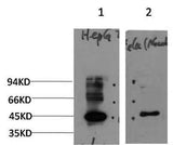
| Specificity | This antibody detects endogenous levels of Aurora A at Human, Mouse,Rat |
|---|---|
| Validation evidence captions | Western blot analysis of 1)HepG2 Cell, 2) Hela Cells treated with Nocodazole using Rabbit pAb diluted at 1:2,000. | Immunohistochemical analysis of paraffin-embedded human Squamous cell carcinoma of lung. 1, Antibody was diluted at 1:200(4° overnight). 2, Tris-EDTA,pH9.0 was used for antigen retrieval. 3,Secondary antibody was diluted at 1:200(room temperature, 45min). |
Key Facts
| Target | Aurora A |
| Also known as | Aurora kinase A (EC 2.7.11.1) (Aurora 2) (Aurora/IPL1-related kinase 1) (ARK-1) (Aurora-related kinase 1) (hARK1) (Breast tumor-amplified kinase) (Serine/threonine-protein kinase 15) (Serine/threonine-protein kinase 6) (Serine/threonine-protein kinase aurora-A) |
| Concentration | 1 mg/ml |
| Working concentration | IHC-p1:50-200 ,WB 1:1000-2000 |
| Species reactivity | Human; Mouse;Rat |
| Observed band | 46kD |
| Cellular localization | Cytoplasm, cytoskeleton, microtubule organizing center, centrosome . Cytoplasm, cytoskeleton, spindle pole . Cytoplasm, cytoskeleton, cilium basal body . Cytoplasm, cytoskeleton, microtubule organizing center, centrosome, centriole . Cell projection, neuron projection . Detected at the neurite hillock in developing neurons (By similarity). Localizes at the centrosome in mitotic cells from early prophase until telophase, but also localizes to the spindle pole MTs from prophase to anaphase (PubMed:9606188, PubMed:17229885, PubMed:21225229). Colocalized with SIRT2 at centrosome (PubMed:22014574). Moves to the midbody during both telophase and cytokinesis (PubMed:17726514). Associates with both the pericentriolar material (PCM) and centrioles (PubMed:22014574). The localization to the spindle |
| Purity | The antibody was affinity-purified from rabbit serum by affinity-chromatography using specific immunogen. |
| Form / buffer | Liquid in PBS containing 50% glycerol, 0.5% BSA and 0.20% sodium azide. |
| Research area | >>Oocyte meiosis;>>Progesterone-mediated oocyte maturation |
| Host species | Rabbit |
| Clonality | Polyclonal |
| Isotype | IgG |
| Conjugation | Unconjugated |
| Tested applications |
WBIHC |
| Dilution range | IHC-p1:50-200 ,WB 1:1000-2000 |
| Immunogen | Synthesized peptide derived from human Aurora A AA range: 65-115 |
| Purification | The antibody was affinity-purified from rabbit serum by affinity-chromatography using specific immunogen. |
Reactivity & Application Validation
3 speciess| Species | WB | IHC | IF | ELISA | IP | FCM | CHIP |
|---|---|---|---|---|---|---|---|
| Human | |||||||
| Mouse | |||||||
| Rat |
| Species | Dilution | Notes |
|---|---|---|
| Human | IHC-p1:50-200 | — |
| Mouse | — | — |
| Rat | — | — |
| Species | Dilution | Notes |
|---|
Specifications
Storage & Stability
Compliance & Certifications
Manufactured under ISO 9001:2015 quality management standards.
Not intended for diagnostic or therapeutic use.