53BP1 rabbit pAb
53BP1 rabbit pAb from ELK Biotechnology Read more ›

Product Description
53BP1 rabbit pAb is an ELK Biotechnology antibody for research workflows involving 53BP1.
Background
function:May have a role in checkpoint signaling during mitosis (By similarity). Enhances TP53-mediated transcriptional activation. Plays a role in the response to DNA damage.,PTM:Asymmetrically dimethylated on Arg residues by PRMT1. Methylation is required for DNA binding.,PTM:Phosphorylated at basal level in the absence of DNA damage. Hyper-phosphorylated in an ATM-dependent manner in response to DNA damage induced by ionizing radiation. Hyper-phosphorylated in an ATR-dependent manner in response to DNA damage induced by UV irradiation.,similarity:Contains 2 BRCT domains.,subcellular location:Associated with kinetochores. Both nuclear and cytoplasmic in some cells. Recruited to sites of DNA damage, such as double stand breaks. Methylation of histone H4 at 'Lys-20' is required for efficient localization to double strand breaks.,subunit:Interacts with IFI202A (By similarity). Binds to the central domain of TP53/p53. May form homo-oligomers. Interacts with DCLRE1C. Interacts with histone H2AFX and this requires phosphorylation of H2AFX on 'Ser-139'. Interacts with histone H4 that has been dimethylated at 'Lys-20'. Has low affinity for histone H4 containing monomethylated 'Lys-20'. Does not bind histone H4 containing unmethylated or trimethylated 'Lys-20'. Has low affinity for histone H3 that has been dimethylated on 'Lys-79'. Has very low affinity for histone H3 that has been monomethylated on 'Lys-79' (in vitro). Does not bind unmethylated histone H3.,
Additional antibody information
| Specificity | 53BP1 Polyclonal Antibody detects endogenous levels of 53BP1 protein. |
|---|---|
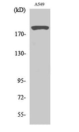
| Validation evidence captions | Western Blot analysis of various cells using 53BP1 Polyclonal Antibody diluted at 1:2000 | Immunofluorescence analysis of HeLa cells, using 53BP1 Antibody. The picture on the right is blocked with the synthesized peptide. | Western blot analysis of lysates from A549 cells, using 53BP1 Antibody. The lane on the right is blocked with the synthesized peptide. | Immunohistochemical analysis of paraffin-embedded human tonsil. 1, Antibody was diluted at 1:200(4° overnight). 2, Tris-EDTA,pH9.0 was used for antigen retrieval. 3,Secondary antibody was diluted at 1:200(room temperature, 45min). |
Key Facts
| Target | 53BP1 |
| Also known as | TP53BP1; Tumor suppressor p53-binding protein 1; 53BP1; p53-binding protein 1; p53BP1 |
| Concentration | 1 mg/ml |
| Working concentration | Western Blot: 1/500 - 1/2000. Immunohistochemistry: 1/100 - 1/300. Immunofluorescence: 1/200 - 1/1000. ELISA: 1/10000. Not yet tested in other applications. |
| Species reactivity | Human;Mouse;Rat |
| Observed band | 213kD |
| Cellular localization | Nucleus . Chromosome . Chromosome, centromere, kinetochore . Localizes to the nucleus in absence of DNA damage (PubMed:28241136). Following DNA damage, recruited to sites of DNA damage, such as double stand breaks (DSBs): recognizes and binds histone H2A monoubiquitinated at 'Lys-15' (H2AK15Ub) and histone H4 dimethylated at 'Lys-20' (H4K20me2), two histone marks that are present at DSBs sites (PubMed:23333306, PubMed:23760478, PubMed:24703952, PubMed:28241136, PubMed:17190600). Associated with kinetochores during mitosis (By similarity). . |
| Purity | The antibody was affinity-purified from rabbit antiserum by affinity-chromatography using epitope-specific immunogen. |
| Form / buffer | Liquid in PBS containing 50% glycerol, 0.5% BSA and 0.02% sodium azide. |
| Research area | >>NOD-like receptor signaling pathway |
| Host species | Rabbit |
| Clonality | Polyclonal |
| Isotype | IgG |
| Conjugation | Unconjugated |
| Tested applications |
WBIHCIFELISA |
| Dilution range | Western Blot: 1/500 - 1/2000. Immunohistochemistry: 1/100 - 1/300. Immunofluorescence: 1/200 - 1/1000. ELISA: 1/10000. Not yet tested in other applications. |
| Immunogen | The antiserum was produced against synthesized peptide derived from human 53BP1. AA range:1-50 |
| Purification | The antibody was affinity-purified from rabbit antiserum by affinity-chromatography using epitope-specific immunogen. |
Reactivity & Application Validation
3 speciess| Species | WB | IHC | IF | ELISA | IP | FCM | CHIP |
|---|---|---|---|---|---|---|---|
| Human | |||||||
| Mouse | |||||||
| Rat |
| Species | Dilution | Notes |
|---|---|---|
| Human | Western Blot: 1/500 - 1/2000. Immunohistochemistry: 1/100 - 1/300. Immunofluorescence: 1/200 - 1/1000. ELISA: 1/10000. Not yet tested in other applications. | — |
| Mouse | — | — |
| Rat | — | — |
| Species | Dilution | Notes |
|---|
| Species | Dilution | Notes |
|---|
| Species | Dilution | Notes |
|---|
Specifications
Storage & Stability
Compliance & Certifications
Manufactured under ISO 9001:2015 quality management standards.
Not intended for diagnostic or therapeutic use.