Smad2 (phospho Ser467) rabbit pAb
Smad2 (phospho Ser467) rabbit pAb from ELK Biotechnology Read more ›

Product Description
Smad2 (phospho Ser467) rabbit pAb is an ELK Biotechnology antibody for research workflows involving Smad2.
Background
The protein encoded by this gene belongs to the SMAD, a family of proteins similar to the gene products of the Drosophila gene 'mothers against decapentaplegic' (Mad) and the C. elegans gene Sma. SMAD proteins are signal transducers and transcriptional modulators that mediate multiple signaling pathways. This protein mediates the signal of the transforming growth factor (TGF)-beta, and thus regulates multiple cellular processes, such as cell proliferation, apoptosis, and differentiation. This protein is recruited to the TGF-beta receptors through its interaction with the SMAD anchor for receptor activation (SARA) protein. In response to TGF-beta signal, this protein is phosphorylated by the TGF-beta receptors. The phosphorylation induces the dissociation of this protein with SARA and the association with the family member SMAD4. The association with SMAD4 is important for the translocation
Additional antibody information
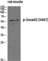
| Specificity | Phospho-Smad2 (S467) Polyclonal Antibody detects endogenous levels of Smad2 protein only when phosphorylated at S467. |
|---|---|
| Validation evidence captions | Western Blot analysis of various cells using Phospho-Smad2 (S467) Polyclonal Antibody diluted at 1:500 | Western Blot analysis of HepG2 cells using Phospho-Smad2 (S467) Polyclonal Antibody diluted at 1:500 | Enzyme-Linked Immunosorbent Assay (Phospho-ELISA) for Immunogen Phosphopeptide (Phospho-left) and Non-Phosphopeptide (Phospho-right), using Smad2 (Phospho-Ser467) Antibody | Immunohistochemistry analysis of paraffin-embedded human breast carcinoma, using Smad2 (Phospho-Ser467) Antibody. The picture on the right is blocked with the phospho peptide. |
Key Facts
| Target | Smad2 |
| Also known as | SMAD2; MADH2; MADR2; Mothers against decapentaplegic homolog 2; MAD homolog 2; Mothers against DPP homolog 2; JV18-1; Mad-related protein 2; hMAD-2; SMAD family member 2; SMAD 2; Smad2; hSMAD2 |
| Concentration | 1 mg/ml |
| Working concentration | Western Blot: 1/500 - 1/2000. Immunohistochemistry: 1/100 - 1/300. ELISA: 1/5000. Not yet tested in other applications. |
| Species reactivity | Human;Mouse;Rat |
| Observed band | 58kD |
| Cellular localization | Cytoplasm . Nucleus . Cytoplasmic and nuclear in the absence of TGF-beta. On TGF-beta stimulation, migrates to the nucleus when complexed with SMAD4 (PubMed:9865696, PubMed:21145499). On dephosphorylation by phosphatase PPM1A, released from the SMAD2/SMAD4 complex, and exported out of the nucleus by interaction with RANBP1 (PubMed:16751101, PubMed:19289081). Localized mainly to the nucleus in the early stages of embryo development with expression becoming evident in the cytoplasm at the blastocyst and epiblast stages (By similarity). . |
| Purity | The antibody was affinity-purified from rabbit antiserum by affinity-chromatography using epitope-specific immunogen. |
| Form / buffer | Liquid in PBS containing 50% glycerol, 0.5% BSA and 0.02% sodium azide. |
| Research area | >>Cell cycle;>>Endocytosis;>>Cellular senescence;>>TGF-beta signaling pathway;>>Apelin signaling pathway;>>Hippo signaling pathway;>>Signaling pathways regulating pluripotency of stem cells;>>Th17 cell differentiation;>>Relaxin signaling pathway;>>AGE-RAGE signaling pathway in diabetic complications;>>Chagas disease;>>Human T-cell leukemia virus 1 infection;>>Pathways in cancer;>>Proteoglycans in cancer;>>Colorectal cancer;>>Pancreatic cancer;>>Hepatocellular carcinoma;>>Gastric cancer;>>Inflammatory bowel disease;>>Diabetic cardiomyopathy |
| Host species | Rabbit |
| Clonality | Polyclonal |
| Isotype | IgG |
| Conjugation | Unconjugated |
| Tested applications |
WBIHCIFELISA |
| Dilution range | Western Blot: 1/500 - 1/2000. Immunohistochemistry: 1/100 - 1/300. ELISA: 1/5000. Not yet tested in other applications. |
| Immunogen | The antiserum was produced against synthesized peptide derived from human Smad2 around the phosphorylation site of Ser467. AA range:418-467 |
| Purification | The antibody was affinity-purified from rabbit antiserum by affinity-chromatography using epitope-specific immunogen. |
Reactivity & Application Validation
3 speciess| Species | WB | IHC | IF | ELISA | IP | FCM | CHIP |
|---|---|---|---|---|---|---|---|
| Human | |||||||
| Mouse | |||||||
| Rat |
| Species | Dilution | Notes |
|---|---|---|
| Human | Western Blot: 1/500 - 1/2000. Immunohistochemistry: 1/100 - 1/300. ELISA: 1/5000. Not yet tested in other applications. | — |
| Mouse | — | — |
| Rat | — | — |
| Species | Dilution | Notes |
|---|
| Species | Dilution | Notes |
|---|
| Species | Dilution | Notes |
|---|
Specifications
Storage & Stability
Compliance & Certifications
Manufactured under ISO 9001:2015 quality management standards.
Not intended for diagnostic or therapeutic use.