RP1 rabbit pAb
RP1 rabbit pAb from ELK Biotechnology Read more ›

Product Description
RP1 rabbit pAb is an ELK Biotechnology antibody for research workflows involving RP1.
Background
This gene encodes a member of the doublecortin family. The protein encoded by this gene contains two doublecortin domains, which bind microtubules and regulate microtubule polymerization. The encoded protein is a photoreceptor microtubule-associated protein and is required for correct stacking of outer segment disc. This protein and the RP1L1 protein, another retinal-specific protein, play essential and synergistic roles in affecting photosensitivity and outer segment morphogenesis of rod photoreceptors. Because of its response to in vivo retinal oxygen levels, this protein was initially named ORP1 (oxygen-regulated protein-1). This protein was subsequently designated RP1 (retinitis pigmentosa 1) when it was found that mutations in this gene cause autosomal dominant retinitis pigmentosa. Mutations in this gene also cause autosomal recessive retinitis pigmentosa. Transcript variants resulted from an alternative promoter and alternative splicings have been found, which overlap the current reference sequence and has several exons upstream and downstream of the current reference sequence. However, the biological validity and full-length nature of some variants cannot be determined at this time.[provided by RefSeq, Sep 2010],
Additional antibody information
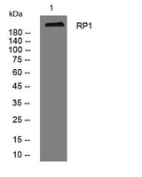
| Specificity | This antibody detects endogenous levels of RP1 at Human/Mouse |
|---|---|
| Validation evidence captions | Western blot analysis of lysates from 293T cells, primary antibody was diluted at 1:1000, 4°over night | Immunohistochemical analysis of paraffin-embedded human liver cancer. 1, Antibody was diluted at 1:200(4° overnight). 2, Tris-EDTA,pH9.0 was used for antigen retrieval. 3,Secondary antibody was diluted at 1:200(room temperature, 45min). |
Key Facts
| Target | RP1 |
| Concentration | 1 mg/ml |
| Working concentration | WB 1:500-2000;IHC-p 1:50-300; ELISA 2000-20000 |
| Species reactivity | Human; Mouse |
| Cellular localization | Cytoplasm, cytoskeleton, cilium axoneme . Cell projection, cilium, photoreceptor outer segment . Specifically localized in the connecting cilia of rod and cone photoreceptors. |
| Purity | The antibody was affinity-purified from rabbit serum by affinity-chromatography using specific immunogen. |
| Form / buffer | Liquid in PBS containing 50% glycerol, 0.5% BSA and 0.02% sodium azide. |
| Research area | Antibody Research |
| Host species | Rabbit |
| Clonality | Polyclonal |
| Isotype | IgG |
| Conjugation | Unconjugated |
| Tested applications |
WBIHCELISA |
| Dilution range | WB 1:500-2000;IHC-p 1:50-300; ELISA 2000-20000 |
| Immunogen | Synthesized peptide derived from human RP1 AA range: 1330-1380 |
| Purification | The antibody was affinity-purified from rabbit serum by affinity-chromatography using specific immunogen. |
Reactivity & Application Validation
2 speciess| Species | WB | IHC | IF | ELISA | IP | FCM | CHIP |
|---|---|---|---|---|---|---|---|
| Human | |||||||
| Mouse |
| Species | Dilution | Notes |
|---|---|---|
| Human | WB 1:500-2000;IHC-p 1:50-300; ELISA 2000-20000 | — |
| Mouse | — | — |
| Species | Dilution | Notes |
|---|
| Species | Dilution | Notes |
|---|
Specifications
Storage & Stability
Compliance & Certifications
Manufactured under ISO 9001:2015 quality management standards.
Not intended for diagnostic or therapeutic use.