NHRF1 rabbit pAb
NHRF1 rabbit pAb from ELK Biotechnology Read more ›

Product Description
NHRF1 rabbit pAb is an ELK Biotechnology antibody for research workflows involving NHRF1.
Background
This gene encodes a sodium/hydrogen exchanger regulatory cofactor. The protein interacts with and regulates various proteins including the cystic fibrosis transmembrane conductance regulator and G-protein coupled receptors such as the beta2-adrenergic receptor and the parathyroid hormone 1 receptor. The protein also interacts with proteins that function as linkers between integral membrane and cytoskeletal proteins. The protein localizes to actin-rich structures including membrane ruffles, microvilli, and filopodia. Mutations in this gene result in hypophosphatemic nephrolithiasis/osteoporosis type 2, and loss of heterozygosity of this gene is implicated in breast cancer.[provided by RefSeq, Sep 2009],
Additional antibody information
| Specificity | NHRF1 Polyclonal Antibody detects endogenous levels of protein. |
|---|---|
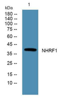
| Validation evidence captions | Western blot analysis of lysates from K562 cells, primary antibody was diluted at 1:1000, 4°over night |
Key Facts
| Target | NHRF1 |
| Concentration | 1 mg/ml |
| Working concentration | WB 1:500-2000 ELISA 1:5000-20000 |
| Species reactivity | Human;Rat;Mouse; |
| Observed band | 39kD |
| Cellular localization | Cytoplasm . Apical cell membrane . Endomembrane system; Peripheral membrane protein. Cell projection, filopodium. Cell projection, ruffle. Cell projection, microvillus. Translocates from the cytoplasm to the apical cell membrane in a PODXL-dependent manner. Colocalizes with CFTR at the midpiece of sperm tail (By similarity). Colocalizes with actin in microvilli-rich apical regions of the syncytiotrophoblast. Found in microvilli, ruffling membrane and filopodia of HeLa cells. Present in lipid rafts of T-cells. . |
| Purity | The antibody was affinity-purified from rabbit antiserum by affinity-chromatography using epitope-specific immunogen. |
| Form / buffer | Liquid in PBS containing 50% glycerol, and 0.02% sodium azide. |
| Research area | >>Tight junction;>>Parathyroid hormone synthesis, secretion and action;>>Pathogenic Escherichia coli infection;>>Human papillomavirus infection |
| Host species | Rabbit |
| Clonality | Polyclonal |
| Isotype | IgG |
| Conjugation | Unconjugated |
| Tested applications |
WBELISA |
| Dilution range | WB 1:500-2000 ELISA 1:5000-20000 |
| Immunogen | Synthesized peptide derived from part region of human protein |
| Purification | The antibody was affinity-purified from rabbit antiserum by affinity-chromatography using epitope-specific immunogen. |
Reactivity & Application Validation
3 speciess| Species | WB | IHC | IF | ELISA | IP | FCM | CHIP |
|---|---|---|---|---|---|---|---|
| Human | |||||||
| Rat | |||||||
| Mouse |
| Species | Dilution | Notes |
|---|---|---|
| Human | WB 1:500-2000 ELISA 1:5000-20000 | — |
| Rat | — | — |
| Mouse | — | — |
| Species | Dilution | Notes |
|---|
Specifications
Storage & Stability
Compliance & Certifications
Manufactured under ISO 9001:2015 quality management standards.
Not intended for diagnostic or therapeutic use.