

Super-resolution confocal microscopy and immunofluorescence imaging, conducted independently.
Viroscope Imaging is a premium scientific imaging agency bridging biology, microscopy, and visual storytelling for life science companies.
The service transforms complex biological data into gallery-grade visual assets while supporting R&D validation, antibody evidence, product launches, investor communication, and institutional campaigns.

Every antibody entering the Viroscope pipeline is subjected to the same non-negotiable sequence. The data produced powers ABMIUM Validated listings.
The antibody is applied to cross-species reactivity, tested in cell-line panels. Establishing baseline performance data and identifying species-specific limitations at the outset.
Varied fixation methods and immunofluorescence protocols are applied systematically to determine optimal conditions and expose failures under non-ideal parameters.
High-end confocal microscopy reveals exact subcellular localisation, producing spatial data that no Western blot or datasheet can replicate.
Reality is documented. A Viroscope negative is published to the same standard as a positive. On ABMIUM, that negative is data you can trust and cite.
Spatial validation uses microscopy to assess whether an antibody produces the expected localisation pattern in the right cellular compartment, under disclosed fixation, staining, imaging, and analysis conditions.
For visual validation, a positive signal is only useful when it is biologically plausible. Nuclear targets should resolve to nuclear compartments, membrane targets should show membrane-associated staining, and cytoplasmic markers should not be accepted without pattern-level review.
This service supports ABMIUM Validated™ standards by converting imaging into structured evidence: target localisation, background behaviour, fixation sensitivity, antibody concentration, cell model, channel settings, and application-specific limitations.
Does the staining pattern match the expected biology for the target and sample model?
Is useful signal distinguishable from non-specific staining, bleed-through, fixation artefact, or imaging noise?
Can the antibody be recommended for the stated visual application, or should limitations be disclosed?
Antibody datasheets are produced by the vendors selling the product. Viroscope Imaging provides objective, third-party validation free from internal manufacturer bias. Our only obligation is to scientific accuracy, which means our validation data carries a weight that in-house data cannot.
When a Viroscope result appears on an ABMIUM product listing, researchers access independently acquired subcellular localisation images, disclosed protocol conditions, and verified species reactivity before purchasing.
Each validated listing on ABMIUM carries independently acquired spatial data, not a reformatted manufacturer claim.
Most antibody validation data only documents what works. Viroscope publishes the full picture. Knowing that an antibody fails under PFA fixation in a specific application is precisely the information a researcher needs before committing budget and samples.
Super-resolution imaging confirmed exclusive nuclear localisation in three human cell lines under PFA and methanol fixation. Cross-reactive in mouse and rat. IF-optimised concentration documented.
PFA fixation produced non-specific cytoplasmic background. Methanol fixation resolved signal to expected nuclear compartment. Application failure documented with exact conditions, a precise protocol warning rather than a product rejection.
A selection of imaging outputs from Viroscope independent validation campaigns. Each image is accompanied by full protocol documentation and is available on the respective ABMIUM product listing.
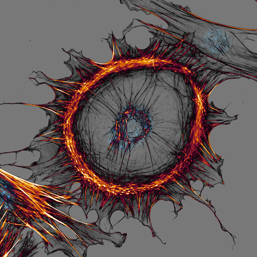
MEF cell cultivated on a poly-L-lysine covered substrate. The actin cytoskeleton, forming a massive central ring and peripheral extensions, is stained in a grey to black to orange Lookup Table. The cell nucleus containing DNA is stained cyan.
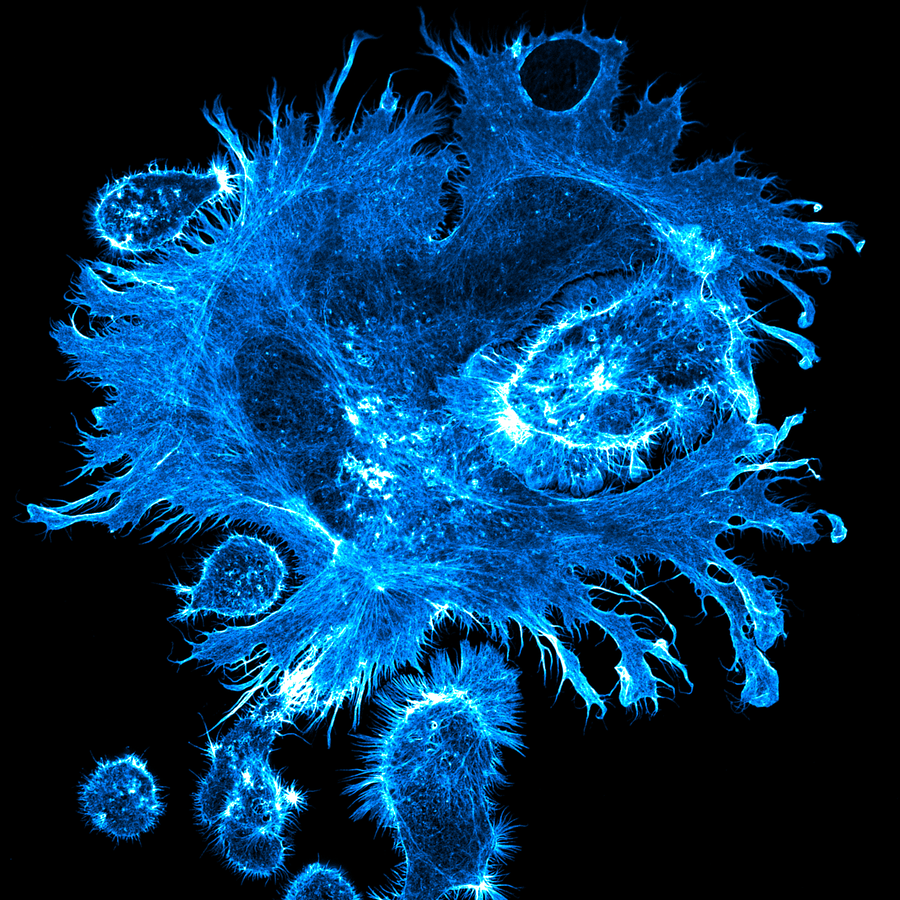
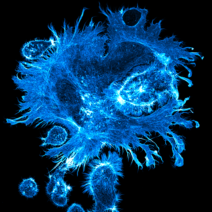
The image shows the actin cytoskeleton in cyan.
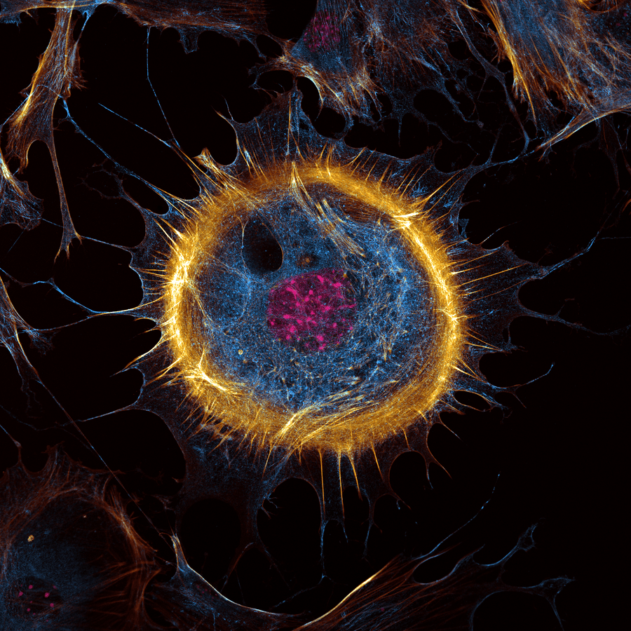
The image shows the actin cytoskeleton in gold, septin cytoskeleton in cyan and DNA in pink.
Use this space to feature awards, publication covers, or recognised scientific imaging highlights alongside the enquiry form.

All fields marked required. We respond within 48 hours.