
p53 (phospho Thr18) rabbit pAb from ELK Biotechnology Read more ›

p53 (phospho Thr18) rabbit pAb is a research-use antibody from ELK Biotechnology for studying p53 in WB, IHC, IF, ELISA workflows with listed reactivity for Human, Mouse, Rat. Specifications and supporting documents below should be reviewed when planning the experiment.
tumor protein p53(TP53) Homo sapiens This gene encodes a tumor suppressor protein containing transcriptional activation, DNA binding, and oligomerization domains. The encoded protein responds to diverse cellular stresses to regulate expression of target genes, thereby inducing cell cycle arrest, apoptosis, senescence, DNA repair, or changes in metabolism. Mutations in this gene are associated with a variety of human cancers, including hereditary cancers such as Li-Fraumeni syndrome. Alternative splicing of this gene and the use of alternate promoters result in multiple transcript variants and isoforms. Additional isoforms have also been shown to result from the use of alternate translation initiation codons (PMIDs: 12032546, 20937277). [provided by RefSeq, Feb 2013],
| Specificity | Phospho-p53 (T18) Polyclonal Antibody detects endogenous levels of p53 protein only when phosphorylated at T18. |
|---|---|
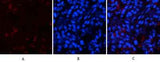
| Validation evidence captions | Immunofluorescence analysis of rat-lung tissue. 1,p53 (phospho Thr18) Polyclonal Antibody(red) was diluted at 1:200(4°C,overnight). 2, Cy3 labled Secondary antibody was diluted at 1:300(room temperature, 50min).3, Picture B: DAPI(blue) 10min. Picture A:Target. Picture B: DAPI. Picture C: merge of A+B | Immunofluorescence analysis of rat-lung tissue. 1,p53 (phospho Thr18) Polyclonal Antibody(red) was diluted at 1:200(4°C,overnight). 2, Cy3 labled Secondary antibody was diluted at 1:300(room temperature, 50min).3, Picture B: DAPI(blue) 10min. Picture A:Target. Picture B: DAPI. Picture C: merge of A+B | Immunohistochemical analysis of paraffin-embedded Human-lung-cancer tissue. 1,p53 (phospho Thr18) Polyclonal Antibody was diluted at 1:200(4°C,overnight). 2, Sodium citrate pH 6.0 was used for antibody retrieval(>98°C,20min). 3,Secondary antibody was diluted at 1:200(room tempeRature, 30min). Negative control was used by secondary antibody only. | Western Blot analysis of various cells using Phospho-p53 (T18) Polyclonal Antibody diluted at 1:2000 |
| Collaborator | ELK Biotechnology |
| Partner model | ES1388 |
| Target | p53 |
| Research areas | >>Endocrine resistance, >>Platinum drug resistance, >>MAPK signaling pathway, >>Sphingolipid signaling pathway, >>Cell cycle, >>p53 signaling pathway, >>Mitophagy - animal, >>PI3K-Akt signaling pathway, >>Apoptosis, >>Longevity regulating pathway, >>Ferroptosis, >>Cellular senescence, >>Wnt signaling pathway, >>Neurotrophin signaling pathway, >>Thyroid hormone signaling pathway, >>Parkinson disease, >>Amyotrophic lateral sclerosis, >>Huntington disease, >>Shigellosis, >>Hepatitis C, >>Hepatitis B, >>Measles, >>Human cytomegalovirus infection, >>Human papillomavirus infection, >>Human T-cell leukemia virus 1 infection, >>Kaposi sarcoma-associated herpesvirus infection, >>Herpes simplex virus 1 infection, >>Epstein-Barr virus infection, >>Pathways in cancer, >>Transcriptional misregulation in cancer, >>Viral carcinogenesis, >>Proteoglycans in cancer, >>MicroRNAs in cancer, >>Colorectal cancer, >>Pancreatic cancer, >>Endometrial cancer, >>Glioma, >>Prostate cancer, >>Thyroid cancer, >>Basal cell carcinoma, >>Melanoma, >>Bladder |
| Reactivity | Human,Mouse,Rat |
| Applications | WB,IHC,IF,ELISA |
| Conjugation | Unconjugated |
| Dilution range | IF: 1:50-200 WB 1:500-2000, IHC 1:50-300 IHC 1:50-300 |
| Host | Rabbit |
| Clonality | Polyclonal |
| Isotype | IgG |
| Immunogen | The antiserum was produced against synthesized peptide derived from human p53 around the phosphorylation site of Thr18. AA range:1-50 |
| Concentration | 1 mg/ml |
| Purification | The antibody was affinity-purified from rabbit antiserum by affinity-chromatography using epitope-specific immunogen. |
| Assay type | Antibody validation/application |
| Expression system | Ubiquitous. Isoforms are expressed in a wide range of normal tissues but in a tissue-dependent manner. Isoform 2 is expressed in most normal tissues but is not detected in brain, lung, prostate, muscle, fetal brain, spinal cord and fetal liver. Isoform 3 is expressed in most normal tissues but is not detected in lung, spleen, testis, fetal brain, spinal cord and fetal liver. Isoform 7 is expressed in most normal tissues but is not detected in prostate, uterus, skeletal muscle and breast. Isoform 8 is detected only in colon, bone marrow, testis, fetal brain and intestine. Isoform 9 is expressed in most normal tissues but is not detected in brain, heart, lung, fetal liver, salivary gland, breast or intestine. |
| Purity | The antibody was affinity-purified from rabbit antiserum by affinity-chromatography using epitope-specific immunogen. |
| Species | WB | IHC | IF | ELISA | IP | FCM | ChIP |
|---|---|---|---|---|---|---|---|
| Human | |||||||
| Mouse | |||||||
| Rat |
| Species | Dilution | Notes |
|---|---|---|
| Human | IF: 1:50-200 WB 1:500-2000 | — |
| Mouse | — | — |
| Rat | — | — |
| Species | Dilution | Notes |
|---|
| Species | Dilution | Notes |
|---|
| Species | Dilution | Notes |
|---|
Collaborator meets ABMIUM quality standards and overall product performance standards have been met.
Quality management certification.
For research applications. Not for diagnostic use.
No customer reviews yet.
